Download
1 / 11
110 likes | 315 Vues
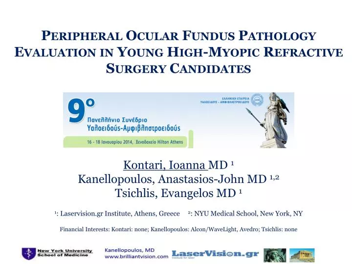
Peripheral Ocular Fundus Pathology Evaluation in Young High-Myopic Refractive Surgery Candidates. Kontari , Ioanna MD 1 Kanellopoulos, Anastasios - John MD 1,2 Tsichlis , Evangelos MD 1 1 : Laservision.gr Institute, Athens, Greece 2 : NYU Medical School, New York, NY

E N D
Peripheral Ocular Fundus Pathology Evaluation in Young High-Myopic Refractive Surgery Candidates Kontari, IoannaMD 1 Kanellopoulos, Anastasios-John MD 1,2 Tsichlis, Evangelos MD 1 1: Laservision.gr Institute, Athens, Greece 2: NYU Medical School, New York, NY Financial Interests: Kontari: none; Kanellopoulos: Alcon/WaveLight, Avedro; Tsichlis: none
to evaluate risk factors in myopic laser refractive surgery candidates, in regard to peripheral fundus pathology. Purpose Setting • a modern ophthalmological surgical center.
Background • Refractive surgery is reported to be a contributing factor in developing retinal lesions and/or RD • Chan CK, Tarasewicz DG, Lin SG. Relation of pre-LASIK and post-LASIK retinal lesions and retinal examination for LASIK eyes. Br J Ophthalmol. 2005 Mar;89(3):299-301. • Luna JD, Artal MN, Reviglio VE, Pelizzari M, Diaz H, Juarez CP. Vitreoretinal alterations following laser in situ keratomileusis: clinical and experimental studies. Graefes Arch ClinExpOphthalmol. 2001 Jul;239(6):416-23. • Rodriguez A, Camacho H. Retinal detachment after refractive surgery for myopia. Retina. 1992;12(3 Suppl):S46-50.
Methods: 6 months, 122 patients • Inclusion criteria: • refractive surgery candidates, age 18 – 35 years • myopia more than -5 D • Reported RD risk: -1 to -3 D, 4× emmetropia > -3 D, 10× emmetropia • Reported RD incidence for myopia more than -6 D, 3.2% - compare to 0.7% for emmetropia • no previous history (trauma, ocular surgery, ocular pathology).
Methods • Anterior Segment Exam protocol: • Visual Acuity (best-corrected distance, uncorrected distance), • pupillometry (photopic and mesopic), • manifest and cycloplegic refraction, • Placido topography, • novel multicolored-spot reflection topography, • Scheimpflug topometry.
Methods • Posterior Segment Exam protocol: • fundus camera documentation • dilated fundus examination with 90° lensand • peripheral fundus examination with Goldmann(3-mirror) lens • Findings were video recorded for future objective documentation and re-evaluation.
Myopia refraction: preoperative refraction -7.5±3.4 (-5 to -18.5) D. 14 eyes (11.4%) were presented with Syneresis (prevalence reported to be 20% in young populace 14-18 years of age), PVD (posterior vitreous detachment), andperipheral fundus lesions such as horseshoe tear without detachment, operculated break without detachment, round hole without detachment lattice degeneration, white without pressure Results:65 Female, 57 Male, Age: 25±5.7 (18 to 35) years
Lattice degeneration • 6-8% in normal population, 50% bilateral • 64-83% RD on the basis of lattice degeneration • 1.4% RD at 10 years* • 423 eyes with lattice degeneration followed for 10 years, developed 0.7% RD* • Prophylactic laser coagulation depends on the fellow eye • * Byer Norman E. Long-term natural history of lattice degeneration of the retina. Ophthalmology, 1989 Sep;96(9):1396-401
Examples The same patient with a superior peripheral retinal tear. Small, layered preretinalhemorrhage at inferior vitreous base in a patient with floaters and an acute PVD. Laser demarcation surrounding an area of subretinal fluid. A bridging vessel over a retinal tear with subretinal fluid.
Conclusions • The prevalence of peripheral fundus pathology in young Greeks with high myopia was increased than previously thought, and perhaps has evaded attention in the past. • Detailed, complete fundus periphery examination is recommended, not just to high-myopic patients, but also to all patients presented for routine screening.
Thank you www.laservision.gr