Download
1 / 44
520 likes | 906 Vues
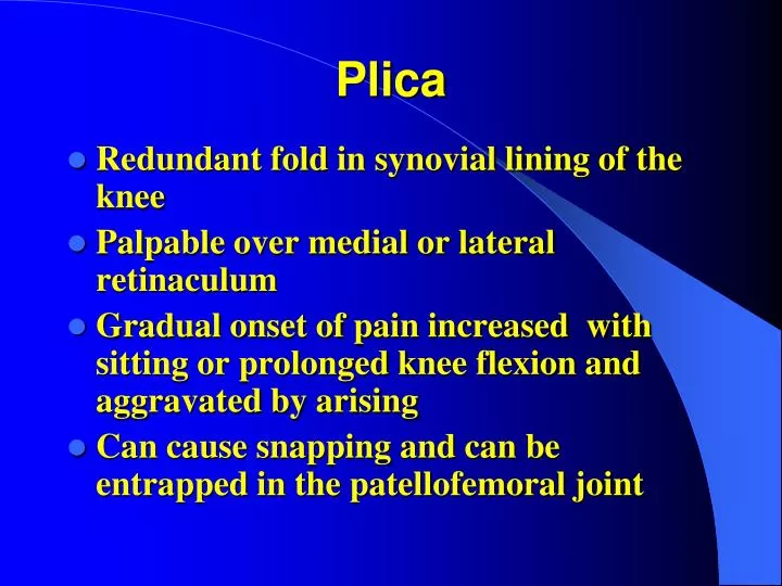
Plica. Redundant fold in synovial lining of the knee Palpable over medial or lateral retinaculum Gradual onset of pain increased with sitting or prolonged knee flexion and aggravated by arising Can cause snapping and can be entrapped in the patellofemoral joint . Plica. Treatment

E N D
Plica • Redundant fold in synovial lining of the knee • Palpable over medial or lateral retinaculum • Gradual onset of pain increased with sitting or prolonged knee flexion and aggravated by arising • Can cause snapping and can be entrapped in the patellofemoral joint
Plica Treatment • Cortisone injection may help • Surgery may be required for excision of the thickened band of tissue
Plica Synovial plica Patella Plica Trochlea groove
Anterior Cruciate Ligament Injuries • Incidence is 0.3-0.38 per 1000 per year and increasing • Usually sports related (football, soccer, skiing) • Estimated 3 in 100,000 have tibial spine avulsions • Skeletally immature patients account for 3-4% of all ACL injuries
Anterior Cruciate Ligament Injuries • Bony avulsion of tibial spine insertion occurs more commonly found in preadolescent children • Intrasubstance tears are more commonly seen in adolescents • MOI is usually hyperextension, sudden deceleration, or a valgus rotational force with a stationary foot
Isolated ACL Tears • Preadolescent children best treated with activity modification and observation • Repair of ligament has high failure rate • Recommendation for children with > 1year left of growth remaining not to have bone tunnels for reconstruction (some controversy) • Adolescents are treated as adults
Anterior Cruciate Ligament Injuries Natural History • Preadolescent –not well known due to low #’s • Adolescents- similar to young adults • 33-86% reports episodes of “giving way” when treated nonoperatively
Anterior Cruciate Ligament Injuries Natural History • Activity, not age is primary factor • Adolescents are usually very active and they will have a higher rate of failure with conservative treatment • Increase risk of meniscal damage if treated conservatively
Tibial Spine Avulsion Fractures • Type I-minimally displaced • Type II-posterior hinge but still attached to the tibial epiphysis • Type III- fx is displaced Type I Type II Type III
Tibial Spine Avulsion Fractures Treatment • Type I – usually casting • Type II – closed reduction/cast • Type III – ORIF with screw/wire/sutures
Mensical Injuries Symptoms • Pain • Effusion • Snapping • Giving way • Intermittent locking • Locked knee
Meniscal Injuries Outcomes • Complete meniscectomies results are very poor • 60% unsatisfactory at 7 years • Preservation is critical • 80-90% have favorable outcomes with repair
Meniscal Injuries Blood supply to meniscus Repairable zone Meniscal tear
Patellar Dislocation Twisting injury Collision May not know patella dislocated Immediate swelling Can’t play
Patellar Dislocation Almost always lateral Younger age at initial dislocation, increased risk of recurrent dislocation Often reduce spontaneously with knee extension and present with hemarthrosis Immobilize in extension for 4 weeks
Patellar Dislocation Predisposing factors to recurrence- ligamentous laxity, increased genu valgum, torsional malalignment Consider surgical treatment for recurrent dislocation/subluxation if fail extensive rehabilitation/exercises
Other LE Injuries Bone Bruise Shin Splints Stress Fractures Nerve Entrapements Severe’s Disease Subungual Hematoma’s
Bone bruise Collision Fall Non-contact twist Xrays usually normal dx by mri Tx rest,nwb time
Shin Splints • Exercise induced pain along the anteriomedial tibia • Encompasses a spectrum of disorders- posterior tibial tendonitis, periostitis, and can lead to a stress fracture • Usually a result of training errors or change in quality and quantity of running
Shin Pain Differential Diagnosis • Shin Splints • Chronic Exertional Compartment Syndrome • Nerve Entrapment Syndromes • Stress Fractures
Shin Splints Treatment • Ice massage • NSAID’s • Decrease running and jumping • Correct training errors • Strengthening • Continue with cardio/vascular training
Stress Fractures • Most commonly results from training errors (change in surfaces or shoes) • Need high index of suspicion • Difficult clinically to distinguish from overuse type syndromes • May be seen on plain X-rays but Tc-99m bone scan may be needed • MRI
Stress Fractures • Increasingly more frequent in recreational and competitive athletes • About 10% of sport injuries • Common with preexisting conditions with decreased bone mass, bone mineralization [nutritional disorders (rickets), systemic disorders (DM, RA)]
Stress Fractures Treatment • Activity modification [you need to keep these athletes in shape (swim, bike, weight train, etc.)] • Rest- (it takes time) • Immobilization rarely needed
Nerve Entrapment Syndromes • Occur secondarily to problems such as lower extremity edema, compression syndrome, bone impingement and joint instability • Nerve problems are mostly functional: that is the nerve is entrapped only during athletic activity
Nerve Entrapment Syndromes • Superficial peroneal n.-pain and numbness over distal calf & dorsum of foot & ankle • Deep peroneal n.- pain at dorsum of foot with pain and numbness at 1st web • Sural n.-numbness at lateral heel and foot • Posterior tibial n.-tarsal tunnel symptoms
Sever’s Disease • Self limiting apohysitis of the oscalcis at the insertion site of the Achilles tendon • Pain over post. oscalcis • Aggravated by activity • Recent growth spurt
Sever’s Disease Increased density and partial fragmentation of the calcaneal apophysis
Sever’s Disease • Most commonly seen in 6 to 10 year old males • Treatment is symptomatic • Mild restriction of activities • Ice • Heel lifts • Achilles stretches • Arch supports
Subungual Hematoma Think open fracture
Injuries About The Shoulder • Little Leaguer’s Shoulder • Shoulder instability • Recurrent dislocations • Clavicle fractures • A-C separations
Little Leaguer’s Shoulder • Pain due to a stress fracture of the humeral physis • Overuse injury • Rest usually takes care of the problem
Little Leaguer’s Shoulder • Treatment is PREVENTION • Restrict # of pitches and innings pitched • Mandatory days of rest • Show proper technique ( no side arm) and do not attempt techniques beyond the current skeletal maturity (curve balls)
Little Leaguer’s Shoulder May help to limit the amount of pitching in the game but doesn’t control practice or at home
Shoulder Instability • Commonly seen in young swimmers and throwing athletes (also volleyball & tennis) • Overuse syndrome • Pain is exacerbated by repetitive overhead activities that cause a stretching of the anterior capsule and musculature of the shoulder
Shoulder Instability • Commonly “loose jointed” • Multiple-joint laxity – check the knees, elbows, MCP joints, thumb to flexor surface of forearm • Check skin hyperelasticity (Marfan’s?) • Usually atraumatic instability • Voluntary instability can be associated with psychologic instability
Shoulder Instability • Physical exam may also show rotator cuff and scapular weakness and apprehension • Multifactorial causes • Motor imbalance • Scapular weakness • Capsular laxity • RARELY IS THIS ROTATOR CUFF DISEASE
Shoulder Instability Treatment • Physical therapy- includes stretching posterior capsule and strengthening anterior musculature, rotator cuff, and the muscles of the scapula ( usually up to 6 months) • Surgical - capsular shrinking has promise for failures at therapy , capsular plication
Recurrent Dislocations • Initial traumatic dislocation • Can lead to long term disability • Age is important fact for recurrent dislocations • Over 50 years of age– very few • Children and adolescents- 25-50% • 90% of young athletes have associated Bankart lesions (avulsion of labrum-ligament complex)
Recurrent Dislocations Treatment • Surgical • Open • Arthroscopically- higher failure rate for athletes under 20-years of age?