Download
1 / 93
990 likes | 1.56k Vues
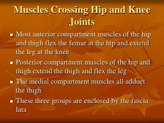
4.1 Joints and Muscles. What would life be like without Joints Move a joint that you use often How do different joints move. Essential Question. 1. What role do joints play in the human body?

E N D
What would life be like without Joints Move a joint that you use often How do different joints move
EssentialQuestion 1. What role do joints play in the human body? Joints are the places where two bones meet and allow movement & flexibility and provides support to the human skeleton. 2. How are joints classified by both structure and function? Functionally, joints are classified by how much motion they allow. Structurally, joints are classified as fibrous, cartilaginous, or synovial.
Joint Classification • Immovable/Fibrous • Do not move—EX: joints in dome of skull and between teeth and jawbone • Partially Moveable/Cartilaginous • Move little—linked by cartilage—EX: vertebrae in spine • Immovable joints and slightly movable joints are restricted mainly to the axial skeleton where protection and stability are key
Joint Classification • Freely Moveable/Synovial • Move in many directions— found at the hip, shoulders, elbows, knees, wrists, and ankles —filled with synovial fluid (acts as lubricant)—these joints have synovial cavities • Freely movable joints are found on the appendicular skeleton and permit flexibility in the limbs.
Activity 4.1.1 Bones, Joints, Action! • Obtain a body system graphic organizer (skeletal view) • Research the six main types of synovial joints • Complete activity through question 6
Essential Question 3. What are the different types of synovial joints? • Pivot joint • Ball-and-Socket joint • Saddle joint • Condyloid (Ellipsoidal) joint • Hinge joint • Plane (Planar or Gliding) joint
Activity 4.1.1 Bones, Joints, Action! • http://www.youtube.com/watch?v=9fZkne0GE9g- cow elbow dissection • Create groups of 3 or 4 • Obtain Gloves, Goggles and Cow Elbow • Look for the movement of the joint, cartilage, tendons and ligaments • Complete the rest of Activity 4.1.1 and Conclusion Questions
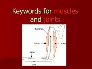
Activity 4.1.1 Bones, Joints, Action! • How are cow elbows and human elbows similar and different? • In the cow joint, the ulna and the radius are fused; whereas in the human, they are two separate bones • Human elbow joint allows for rotation and overall dexterity (not needed by the cow) • What type of synovial joint was modeled by cow elbow?
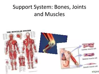
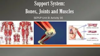
Connective Tissue • Connective tissue protects, supports, and binds together other body tissues. • Connective tissue is made up of different types of cells in varying amounts of a nonliving substance around the cells, called the matrix. • Fibrous connective tissue which is found in tendons and ligaments. Fibrous connective tissue is composed of large amounts of closely packed collagenous fibers. • Cartilage is a form of fibrous connective tissue that is composed of closely packed collagenous fibers in a rubbery gelatinous substance called chondrin.
Essential Question • 4. What role do cartilage, tendons, and ligaments play at a joint? • Cartilage - Cushions/protects bones where they meet and rub against each other. The cartilage found in joints is hyaline cartilage—the same kind found in a fetal skeleton & it’s referred to as articular cartilage where it attaches to articular bone surfaces. • Tendons - Fibrous tissue that connects muscles to bones • Ligaments -Fibrous straps that fasten bones to other bones
Motion with the Cow Elbow • How did your cow elbow move? • Think about the type of synovial joint • How do human elbows move?
Flexibility What makes our bodies flexible? Joints
Joints • Joints with a large range of motion has limited stability • Joint with limited mobility, such as the sutures in the skull, have great stability • Move your hip and shoulder and describe the range of motion of each joint • Joints make up for a lack of stability by the addition of muscle • Joints that are very stable and produce little movement are assisted by limited amounts of muscle • Joints that are very flexible, but offer little stability are surrounded by large amounts of muscle
Describing Motion • How do we describe the motions of a joint ? • Bending • Flexing • Scientists and medical professionals use precise terms to describe the direction of motion as well as the relationship of one body part to another • Depression and Elevation (Make this Motion)
Elevation and Depression Movement Elevation Depression
Activity 4.1.2: Range of Motion • In groups of 3 research the following Terms and Document on your Body Organizers (Complete Steps 1 & 2) • Depression and elevation • Rotation and circumduction • Flexion and extension (and hyperextension) • Abduction and adduction • Plantar flexion and dorsiflexion • When you know all the Motion as a group demonstrate them to your teacher
Essential Question • 5. What terms describe the path of movement at a joint?
Essential Question • 6. What is range of motion? • Range of motion is the range through which a joint can be moved & can be measured using a goniometer to determine angles.
Essential Question • 7. How do you measure the range of motion of a particular joint movement? • Each specific joint has a normal range of motion that is expressed in degrees. • Devices to measure range of motion in the joints of the body include the goniometer and inclinometer which use a stationary arm, protractor, fulcrum, and movement arm to measure angle from axis of the joint. • As measurement results will vary by the degree of resistance, two levels of range of motion results are recorded in most cases.
Finish Activity 4.1.2: Range of Motion • You will need • Activity 4.1.2 Range of Motion • Activity 4.1.2 Student Resource Sheet - ROM • ROM Schematics • Goniometer • http://www.youtube.com/watch?feature=player_embedded&v=ZUF7tpkVAIY- or http://www.youtube.com/watch?v=J_R-igYFj98&feature=player_embedded Instructions on how to use a goniometer
Essential Question • 8. How do bones, muscles and joints work together to enable movement and locomotion for the human body? • Our bones provide support and give our bodies shape, but cannot move on their own. The muscles provide the movement. The joints help attach bones to one another to provide flexibility & allow the muscles to help give the bones a way to move.
Essential Question • 1. How do muscles assist with movement of the body and of substances around the body? • Our muscles are what allow all movement of our bodies (and within our bodies). They help us involuntarily by helping food move down the esophagus and into the stomach (peristalsis) and helping blood move through our bodies (the heart is a muscle). They also help us move our bodies voluntarily from place to place (the muscles in our limbs). Our bodies each have about 650 muscles & are ~ 50% muscle by weight!
With a partner research the following 3 muscle tissues skeletal muscle smooth muscle cardiac muscle Create this table in your Journal View prepared slides Complete Part 1 only Activity 4.2.1 Muscle Rules Part 1
Essential Question • 2. How do the structure and function of the three types of muscle tissue compare? Cardiac- They are striated muscle fibers form the wall of the heart & function involuntarily. Skeletal -They are attached to bone, mostly in the legs, arms, abdomen, chest, neck and face. They are striated muscle fibers (lined under microscope) & attach to bone by a tendon. They hold the skeleton together and give the body shape. They are voluntary (we control them) and contract quickly and powerfully), but they tire easily. Smooth -They are smooth (not striated) & are controlled automatically by our nervous system. They are also called ―involuntary‖ muscles. They make up the walls of the stomach and intestine to help break down and move food. They also line the walls of blood vessels. They take longer to contract than skeletal muscles, but also don’t tire as easily.
Skeletal Muscle • Voluntary – we control the movement • Striated – looks like long fibers • Linked to bones by tendons • Function – to help us move / move our bones
Smooth Muscle • Involuntary Action – controlled by our CNS • Non-striated • Found in arteries, veins, intestines, etc. • Function : Maintain organ dimensions – stretch and recoil
Cardiac Muscle • Involuntary • Striated – but, may be branched which is unlike skeletal muscle. • Found in walls of the heart • Function : To pump the heart! • Highly resistant to fatigue w/lots of mitochondria
Let’s Start By Building a Muscle from Spaghetti Activity 4.2.1 Muscle Rules Part 2 • Pick up one piece of spaghetti. • Each piece of spaghetti will represent one skeletal muscle cell or fiber • Each muscle fiber is enclosed by a delicate membrane called the endomysium. (For the purposes of this activity, the yellow outer coating of the spaghetti represents this membrane.)
Spaghetti Muscle Cont’d • Pick up a handful of spaghetti. This bundle of fibers represents a fascicle. • Each fascicle, however, is covered by a membrane called the perimysium. • Place the bunch of spaghetti on the end of a piece of plastic wrap. • Roll the spaghetti up in the plastic used to represent the perimysium. • Hold up the completed fascicle. • Pull the ends taut, and notice that this tissue has little to no bumps. • These ends represent dense regular connective tissue.
Spaghetti Muscle Cont’d • Fascicles group together to form a skeletal muscle. • Combine your fascicle w/ three other pairs’ to form a whole muscle. • These fascicles are bound together by an even tougher outer membrane called the epimysium. • Wrap the combined fascicles in another piece of plastic wrap. • This layer of wrap will represent the epimysium. • Twist the plastic wrap on each end of the completed muscle. • At the ends of the muscle, the epimysia blend together to form tendons, cordlike structures that attach muscle to bone, cartilage or other connective tissue.
Essential Question 3. How are muscle fibers and membranes organized to form a whole skeletal muscle? The epimysium(“upon muscle”) is the outermost layer of connective tissue. The perimysium (“around muscle”) is made of connective tissue and forms casings for bundles of muscle fibers. The endomysium (“within muscle‖) is connective tissue surrounding each individual muscle fiber. Each fascicle is a small cluster of muscle fibers, with endomysium between the individual fibers. Blood vessels run between the fascicles, bringing the tissue nutrients & removing waste. Nerves also run throughout, controlling the movement of the muscles. Together, the network of nerves and blood vessels are referred to as the plexus
Activity 4.2.1 Muscle Rules Part 3 • Will need your • Manikins • Clay • Lab Journals • We will create a muscle together
Step 1 • Locate the ventral side and use a pencil to place a dot on the lateral and medial side of the radial groove (about halfway up the humerus).
Step 2 • Locate the ulna just below the fold of the elbow. Help the students see the hollowed out area in the antecubital region. Place a pencil dot above this area.
Step 2 Cont’d - Rule 1 • These dots each represent an attachment point for a muscle. • Note that there are at least two attachments (in this case three) and the muscle will cross a joint at the elbow. • This leads us to Muscle Rule #1: Muscles must have at least two attachments and must cross at least one joint.
Step 3 – Brachialis Muscle • Using terra cotta clay, form two balls about the diameter of a nickel. • Rolling the clay between the tabletop and a palm, roll each ball into a long carrot. The total length of the carrots should stretch from the humeral attachment to the ulnar attachment. • Bring the fat part of the carrots together, leaving the tops free (rabbit ears).
Step 4 – Rule 2 • Using your left thumb to represent the humeral attachments and your left middle finger to represent the ulnar attachment, place the left hand on the right arm where the attachments would be. • Make sure to cross the joint. • Pull your forearm towards your heart and watch the position of your fingers. • You should notice that your index finger and thumb are closer together than when you started. • This lead to rule 2: Muscles always “pull” and get shorter.
Step 5 – Rule 3 • Repeat the motion and identify which attachment is “pulling” or moving closer to the other attachment. • The attachment that moves is known as the insertion of the muscle. • The insertion is usually the distal attachment. • The attachment that does not move and pulls the other attachment toward it is referred to as the origin. • The origin is usually the proximal attachment. • This leads to Rule 3: The attachment that moves is known as the insertion and the attachment that remains stationary is known as the origin.
Step 6 • Extend your arms out in front of their bodies. Notice this angle is 180°. • Show the movement again of the muscle you have just built. • This time pay attention to what happens to this angle when the muscle shortens. • Notice that the angle decreases. • Do you remember what we call motion at a joint that decreases the angle between articulating bones? • Flexionand thus a muscle such as this is referred to as a flexor.
Step 7 – Rule 4 • Flex your arms one more time, but stop at the end of the movement. • If muscles only pull, then how can the arm be straightened? • What do we call motion at a joint that increases the angle between articulating bones? • Extension and thus a muscle that controls this movement is referred to as an extensor. Muscles that decrease the angle between ventral surfaces of the body are known as flexors. Muscles that increase the angle between ventral surfaces of the body are known as extensors
Step 8 • Place a pencil dot halfway up the dorsal side of the humerus. • Place another dot just distal of the elbow onto the ulna