Download
1 / 1
10 likes | 369 Vues
No. 061. Female urethral diverticula : evaluation of voiding dysfunction before and after surgery. Wilson Ailsa, Hussain Mahreen , Hamid Rizwan , Ockrim Jeremy, Shah Julian, Greenwell Tamsin University College London Hospitals, United Kingdom. Results

E N D
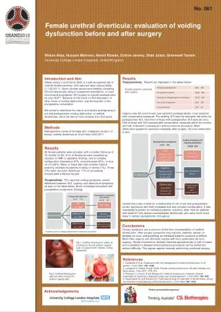
No. 061 Female urethral diverticula: evaluation of voiding dysfunction before and after surgery Wilson Ailsa, HussainMahreen, HamidRizwan, Ockrim Jeremy, Shah Julian, Greenwell Tamsin University College London Hospitals, United Kingdom Results Postoperatively: Results are displayed in the tables below. Urgency was the commonest new symptom postoperatively; most resolved with conservative measures. Pre-existing SUI was the strongest risk factor for postoperative SUI. One third of those with postoperative SUI were de novo. Half of those with SUI resolved with conservative measures within six months, and half underwent a subsequent antiincontinence procedure. Most obstructive symptoms resolved completely after surgery. De novo obstruction is rare. Overall there was a trend for a relationship of risk of pre and postoperative urinary symptoms with both increased size and complex configuration. It was impossible to predict an individual patients’ outcome, other than for patients with small (≤1 cm) simple uncomplicated diverticulae, who were much more likely to remain asymptomatic throughout. Introduction and Aim Stress urinary incontinence (SUI) is a well recognised risk of urethral diverticulectomy, with reported rates varying wildly (1.7-20.3%1,2). Some centres recommend treating coexisting SUI simultaneously (sling or suspension procedure), or even recommend prophylactic SUI surgery to prevent postoperative de novo SUI3,4. Because of this focus on SUI discussion of other forms of voiding dysfunction may be forgotten in the preoperative consultation. We aimed to determine the rate of and factors predisposing to pre and postoperative voiding dysfunction in urethral diverticulae, which we feel is more complex than SUI alone. Overall symptom outcomes after surgery Methods Retrospective review of females who underwent excision of benign urethral diverticula at UCLH from 2000-2011. Results 42 female patients were included, with a median follow up of 18 months (4-96). 81% of diverticula were classified as complex on MRI or operative findings, due to complex configuration (horseshoe 67%, circumferential 22%), or size >3 cm (36%). Many of these also had complex internal anatomy (multiple loculations or ostia) or stones (7%). Three (7%) were recurrent diverticula. 71% of procedures incorporated a Martius fat pad. Preoperatively: 75% reported voiding symptoms, evenly distributed between SUI, urgency and obstructive symptoms as seen in the table below, which correlated somewhat with preoperative urodynamic findings. Conclusions Urinary symptoms are a common at the time of presentation of urethral diverticulum. After surgery symptoms may improve, stabilise, worsen or develop ne novo, and predicting an individual patient’s outcome is difficult. Most often urgency will ultimately resolve with time, particularly de novo urgency. Stress incontinence similarly resolves spontaneously in half of cases, and if persistent a delayed antiincontinence procedure can be performed without difficulty. This argues against routinely performing combined surgery. * 4 patients did not undergo preoperative urodynamic evaluation Fig 1: Urethral diverticulum visible as a fullness in the left anterior vaginal wall, in a patient with irritative voiding symptoms. References 1. Ganabathi K et al. Experience with the management of urethral diverticulum in 63 women. J Urol 1994; 152: 1445. 2. Ljungqvist L, Peeker R and Fall M: Female urethral diverticulum: 26-year followup of a large series. J Urol 2007; 177: 219. 3. Romanzi LJ, Groutz A and Blaivas JG: Urethral diverticulum in women: diverse presentations resulting in diagnostic delay and mismanagement. J Urol2000; 164: 428. 4. Swierzewski SJ and McGuire EJ: Pubovaginal sling for treatment of female stress urinary incontinence complicated by urethral diverticulum. J Urol1993; 149: 1012. Fig 2: Urethral diverticulum exposed after incising the anterior vaginal wall. Poster presentation sponsor Acknowledgements