Download
1 / 1
10 likes | 131 Vues
Integration of Statistical Shape Modeling and Finite Element A nalysis for the Study of Hip Pathology. Sumedha Singla 1,3 ; Andy Anderson 2 ; Ross T. Whitaker 3 ; Jeffrey A. Weiss 1,2,3

E N D
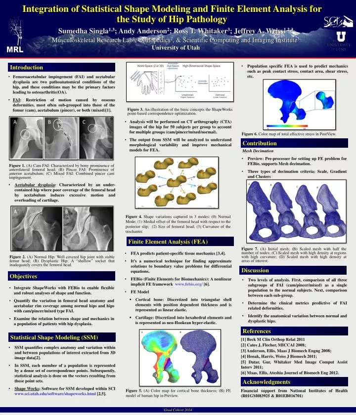
Integration of Statistical Shape Modeling and Finite Element Analysis for the Study of Hip Pathology Sumedha Singla1,3; Andy Anderson2; Ross T. Whitaker3; Jeffrey A. Weiss1,2,3 Musculoskeletal Research Lab1, Orthopedics2, & Scientific Computing and Imaging Institute3University of Utah Introduction • Population specific FEA is used to predict mechanics such as peak contact stress, contact area, shear stress, etc. • Femoroacetabular impingement (FAI) and acetabular dysplasia are two pathoanatomical conditions of the hip, and these conditions may be the primary factors leading to osteoarthritis(OA). • FAI: Restriction of motion caused by osseous deformities,most often sub-grouped into those of the femur (cam), acetabulum (pincer), or both (mixed)[1]. Figure 3. An illustration of the basic concepts the ShapeWorks point-based correspondence optimization. • Analysis will be performed on CT arthrography (CTA) images of the hip for 50 subjects per group to account for multiple groups (cam/pincer/mixed/normal). • The output from SSM will be analyzed to understand morphological variability and improve mechanical models for FEA. Figure 6. Color map of total effective stress in PostView. Contribution • Mesh Decimation • Preview: Pre-processer for setting up FE problem for FEBio, supports Mesh decimation. • Three types of decimation criteria: Scale, Gradient and Clusters Figure 1. (A) Cam FAI: Characterized by bony prominence of anterolateral femoral head; (B) Pincer FAI: Prominence of anterior acetabulum; (C) Mixed FAI: Combined pincer cam impingement. • Acetabular dysplasia: Characterized by an under-contained hip where poor coverage of the femoral head by acetabulum induces excessive motion and overloading of cartilage. Figure 4. Shape variations captured in 3 modes: (0) Normal Mode; (1) Medial offset of the femoral head with respect to the posterior slip; (2) Size of femoral head; (3) Curvature of the trochanter. Finite Element Analysis (FEA) Figure 7. (A) Initial mesh; (B) Scaled mesh with half the number of nodes; (C) Scaled mesh with high density at regions with high curvature; (D) Scaled mesh with high density at areas of interest. • FEA predicts patient-specific tissue mechanics [3,4]. • It’s a numerical technique for finding approximate solutions to boundary value problems for differential equations. • FEBio (Finite Elements for Biomechanics): A nonlinear implicit FE framework www.febio.org/[6]. • FE Model • Cortical bone: Discretized into triangular shell elements with position dependent thickness and is represented as linear elastic. • Cartilage: Discretized into hexahedral elements and is represented as neo-Hookean hyper-elastic. Figure 2. (A) Normal Hip: Well covered hip joint with stable femur head; (B) Dysplastic Hip: A “shallow” socket that inadequately covers the femoral head. Discussion Objectives • Two levels of analysis. First, comparison of all three subgroups of FAI (cam/pincer/mixed) as a single population to the normal subjects. Next, comparison between each sub-group. • Determine the clinical metrics predictive of FAI related deformities. • Identify the anatomical variation between normal and dysplastic hips. • Integrate ShapeWorks with FEBio to enable flexible and robust analyses of shape and function. • Quantify the variation in femoral head anatomy and acetabular rim coverage among normal hips and hips with cam/pincer/mixed type FAI. • Examine the relation between shape and mechanics in a population of patients with hip dysplasia. References Statistical Shape Modeling (SSM) [1] Beck M ClinOrthopRelat 2011 [2] Cates J, Flecher, MICCAI 2008; [3] Anderson, Ellis, Maas J BiomechEngng 2008; [4] Henak, Harris, Weiss J Biomech 2011; [5] Datar, Gur, Whitaker Med Image Comput Assist Interv 2011; [6] Maas, Ellis, AteshiaJournel of BiomechEng 2012. • SSM quantifies complex anatomy and variation within and between populations of interest extracted from 3D image data[2]. • In SSM, each member of a population is represented by a dense set of correspondence points. Subsequently, statistical analysis is done on the vectors resulting from those point sets. • Shape Works: Software for SSM developed within SCI www.sci.utah.edu/software/shapeworks.html [2,5]. Acknowledgments Figure 5. (A) Color map for cortical bone thickness; (B) FE model of human hip in Preview. Financial support from National Institutes of Health (R01GM083925 & R01EB016701) Grad Cohort 2014