Download
1 / 23
301 likes | 919 Vues
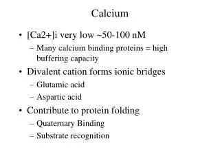
Roles of Calcium. Catalysis Signaling and regulation Muscle contraction Blood clotting Biominerals. Biological Functions of Calcium. Calcium-controlled cellular events. Protein. Location and function. Calmodulin. Cytoplasm, trigger of kinases, etc. Calcineurin.

E N D
Roles of Calcium • Catalysis • Signaling and regulation • Muscle contraction • Blood clotting • Biominerals
Biological Functions of Calcium Calcium-controlled cellular events
Protein Location and function Calmodulin Cytoplasm, trigger of kinases, etc. Calcineurin Cytoplasm, trigger of phosphatases Annexins Internal associated with lipids, trigger C-2 domains Part of several membrane-link enzymes S-100 Internal and External: buffer, messenger, trigger EGF-domains External growth factor but general protein assembly control e.g. fibrillin GLA-domains External, associated with bone Cadherins Cell–cell adhesion Calsequestin Calcium store in reticula ATP-ases Calcium pumps Some classes of calcium proteins in eukaryotes Examples of Calcium-containing proteins B.B.A.1763, 1139 (2006)
Calcium Concentrations Calcium concentrations in cells are quite low while extracellular Ca levels are significantly higher The challenge is to regulate calcium levels and also to be able to change levels rapidly when needed CAM – calmodulin PV – parvalbumin T - troponin ICBP – Ca binding protein The flow into and out of the cells and the various organelles is controlled by individual channel and pump proteins
Calcium Transport Across Membranes & : Ca-ATPase driven transport & : receptor mediated Ca channels & : Na/Ca exchangers & : voltage sensitive Ca channels
Calcium Distribution There are several routes for Ca uptake and for export calcium carrier ATP driven Ca export ATP driven Ca uptake calcium binding proteins Ca/Na antiporter calcium storage Many proteins play important roles in calcium metabolism: calmodulin calcineurin cadherin calsequestrin parvalbumin troponin calpain Ca-ATPase
Protein Activation by Calmodulin Calmodulin can activate different groups of target proteins Calmodulin Kinase Cascade Increase in cellular [Ca(II)] causes Ca binding to CaM CaM binds and activates its target kinase (CaMKIV) Phosphorylation by another kinase (CaMKK) leads to a fully active kinase Phosphorylation of transcription factors leads to activation of transcription CaMKK also phosphorylates a protein kinase (PKB) which leads to inhibition of cell death BioEssays.24, 625 (2002)
Calmodulin structure Calmodulin can bind up to four Ca(II) ions in sets of two each The binding of each pair of metal ions induces changes in the protein conformation that allows it to bind to different target proteins These targets can have very different amino acid sequences, but they do have similar recognition sites for calmodulin Calmodulin binding sites on target proteins
Calmodulin binding The calmodulin domains can wrap around its target protein site The Ca ions are not directly involved in protein-protein contacts, but help to maintain the structural integrity of calmodulin
Calmodulin binding Details of the interactions between calmodulin and a calbindin domain peptide The hydrophobic residues of calmodulin are shown incyan The Met residues of calmodulin are displayed inblue The hydrophobic residues of the calbindin peptide are inbrown Biochemistry45, 738 (2006)
Activation of Calmodulin Kinase Inactive enzyme form autoinhibitory region blocks active site access binding of 4 Ca(II) ions to CaM induces a conformational change that allows CaM to bind and remove the autoinhibitory region This generates the active form of the kinase that can now phosphorylate and activate its protein targets BioEssays.24, 625 (2002)
Binding of Calmodulin to Protein Kinases Since the CaM binding domains of different protein kinases have quite different sequences, Calmodulin can use different binding modes to interact with different protein targets The helical peptides (in blue) from different protein kinases bind in different orientations and interact with different side chain functional groups BioEssays.24, 625 (2002)
Calcineurin Calcineurin (Cn) is a calmodulin-dependent protein phosphatase also known as protein phosphatase 2B. Calcineurin is responsible for activating the transcription of interleukin 2, that stimulates the growth and differentiation of T cell response. In immunosuppressive therapy it is inhibited by cyclosporin and FK506 - drugs known as calcineurin inhibitors. Regulation of Calcineurin Activity The calcineurin phosphatase catalytic domain active site is blocked by an autoinhibitory domain (AID) binding of Ca(II) to CnB causes a conformation change in CnA leading to partial activation this also allows calmodulin (CaM) to bind and remove the AID domain leading to full activity the immunosuppressor FK506 and its binding protein (FKBP) inhibit the enzyme by masking the active site BioEssays.24, 625 (2002)
Calcium bindingtriggered conformational changes The calcium site in the subdomain of a Ca binding protein consisting of a cluster of carboxylate groups Binding of calcium ions induces a conformational change in this loop This local conformational change causes a reorientation of the attached α-helices which exposes a hydrophobic pocket for the binding of the immunosuppressor drugs
Calcineurin complexes Calcineurin complexes with immunosuppressors Complex with cyclosporin A and cyclophilin Complex with FK506 and FK binding protein Another view of the complex with FK506 and FKBP showing the catalytic site and the location of the bound Ca ions P.N.A.S. 99, 13522 (2002)
Role of Calcium in Muscle Contraction • Ca ions are stored bound to calsequestrin • Upon receipt of a nerve or chemical signal, a Ca ion pulse is release through a group of channel proteins • Binding to troponin C triggers a conformation change that causes contraction • Ca ions are removed by binding to parvalbumin • Ca ions are them pumped back into the muscle cells by a Ca-dependent ATPase
Role of Calcium in Blood Clotting Signal amplification by protein cascades The binding of a chemical signal to a cell surface receptor triggers Ca(II) release The binding of Ca activates calpain and stimulates proteases to cleave prothrombin Calpain is responsible for the activation for protein kinases and Protein kinases phosphorylate and activate target proteins TGase triggers the cross-linking of structural proteins
Cell-cell adhesion Adhesion between adjacent cells is important for structural integrity in multicellular organisms A group of calcium binding proteins called cadherins play an important role in cellular adhesion Binding mechanism of cadherins Cadherins are synthesized as inactive precursors with a blocked N-terminus Upon proteolytic activation they exist in an equilibrium between a closed form in which the terminal adhesion arm is docked by intercalating Trp2 into a pocket, and an open form in which the N-terminal is unbound Cell adhesion involves strand exchange to form adhesive dimers with the strands docked into the binding pocket of an adjacent cadherin molecule
Cadherin Dimer structure of human cadherin Three Ca ions bind at the domain interface within each molecule to provide structural rigidity Each interacting molecule inserts its N-terminal peptide into a hydrophobic acceptor site on an adjacent molecule Trp2 of molecule A intercalates between Glu89 and Met92 of molecule B Glu89 of molecule B forms an electrostatic bond to the amino-terminal group of molecule A J. Mol. Biol.373, 401 (2007)
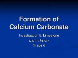
Biominerals Biominerals are composed of different low solubility metal salts that provide structural support
Structure of Biominerals The organic building block for biomineral growth is a fibrous protein called collagen Carboxyl side chains along with phosphorylated lysines provide the ligands to bind calcium ions Calcium ions then bind to phosphate and hydroxyl groups Which, in turn, can bind more calcium ions
Summary • The uptake and export of Ca ions from cells are controlled by channel and pump proteins • Calmodulin activates different groups of protein targets depending on Ca levels • Calcineurin activates interleukin-2 as part of the signalling pathway in the immune system • Cadherins are involved in cellular adhesion • Ca ions play essential roles in muscle contraction, blood clotting, and the formation of biominerals