Download

1 / 70
710 likes | 973 Vues
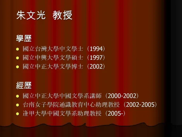
內科專科醫師 職業醫學專科醫師 胸腔暨重症醫學專科醫師 結核病專科醫師. 索任. 簡歷. 現任 2005/5 - 社團法人中華民國防癆協會第一胸腔病防治所所長 2006/1- 行政院衛生署傳染病防治諮詢委員會結核病防治組召集人 2002- 中華民國職業病醫學會理事 1994- 台灣結核病醫學會理事 曾任 2002/7 – 2005/3 行政院衛生署桃園醫院內科師一級主治醫師 2002/2 – 2002/7 行政院衛生署胸腔病院代理院長 2001/8 – 2002/1 行政院衛生署慢性病防治局代理局長

E N D
內科專科醫師職業醫學專科醫師胸腔暨重症醫學專科醫師結核病專科醫師內科專科醫師職業醫學專科醫師胸腔暨重症醫學專科醫師結核病專科醫師 索任 簡歷 • 現任 • 2005/5 - 社團法人中華民國防癆協會第一胸腔病防治所所長 • 2006/1- 行政院衛生署傳染病防治諮詢委員會結核病防治組召集人 • 2002- 中華民國職業病醫學會理事 • 1994- 台灣結核病醫學會理事 • 曾任 • 2002/7 – 2005/3 行政院衛生署桃園醫院內科師一級主治醫師 • 2002/2 – 2002/7 行政院衛生署胸腔病院代理院長 • 2001/8 – 2002/1 行政院衛生署慢性病防治局代理局長 • 1999/7 – 2001/8 行政院衛生署慢性病防治局副局長 • 1997/1 – 1999/6 台北縣慢性病防治所醫師 • 1989/3 – 1997/1 台灣省慢性病防治局技正兼主任 • 1984/4 – 1989/3 台灣省防癆局技正兼主任 • 1983/7 – 1984/4 台灣省防癆局主治醫師 • 1979/7 – 1983/7 台灣省防癆局住院醫師及總住院醫師 1979 台灣大學醫學院醫學系
Tuberculosis and diseases caused by nontuberculosis mycobacteria in Taiwan: Clinical aspect 社團法人中華民國防癆協會 第一胸腔病防治所 索 任 醫 師 solo@tstb.org http://www.tb.org.tw 台北市民權西路104號2樓
The Genus Mycobacterium • Gram-positive, acid-fast, non-spore-forming, straight or slightly curved rods (0.2~0.6 1.0~10m) coccobacillary, filamentous, branched • Pigment in dark or after exposure to light • Aerobic or microaerophilic • Subdivision: rapid growers, slow growers • Large amounts of lipid in cell walls: mycolic acids • Typical species: Mycobacterium tuberculosis
Classification of mycobacterial species commonly cause human disease • Mycobacterium tuberculosis complex • M. leprae • Slowly growing mycobacteria • Photochromogens (Runyon group I) • Scotochromogens (Runyon group II) • Nonchromogens (Runyon group III) • Rapidly growing mycobacteria • (Runyon group IV)
Mycobacterium tuberculosis complex • M. tuberculosis • M. bovis • M. africanum • M. microti • M. canetti M. tb M. kansasii
結核病 –被遺忘了的瘟疫 Tuberculosis 1992
Latent infection vs. disease 感染 發病 感染:會呼吸的人都是結核病的易感宿主 發病:新近感染及免疫力減弱為主要危險因子
如何檢查結核菌感染 • 結核菌素測驗 (不夠精準的檢查) • 結核菌素 tuberculin (Koch,1890) • Mantoux test (1907) • 干擾 – • 宿主的抵抗力, • 卡介苗或其他非結核分枝桿菌(NTM)感染, • 測驗技術和判讀經驗
怎樣檢查肺結核病? 胸部X光檢查 驗痰
TB TB 何時了 潛伏感染 發病 大女兒 69-4-3 F 82-3-13 “5” M+C+ ST N/A 82-12 完治 大兒子 67-11-11 M 95-2-23 “5” M+ 小女兒 71-3-19 F 88-6-28 “3” M+C+ HS 抗藥 89-4 完治 父 39-10-15 M 75-4-7 “5” M+C+ HS 抗藥 76-10 完治
結核病 Class I Exposure Class II 83-4-11 Latent TB Infection 81-3-27 Class III Disease Infectious Non Infectious Death
TB TB 何時了 – 診斷延遲 93/10/12 94/11/30
TB TB 何時了 – 診斷延遲 95/01/12 94/02/23
TB TB 何時了 – 不規則治療 910529 940909
傳染 transmission 化學治療 Chemotherapy 結核病的傳染期= 病人延遲 +醫師延遲 +病人治療管理不當 Doctor’s delay 醫師延誤 Patient’s delay 預防性 Prophylactic 病人延誤 Preventive therapy 治療 treatment 傳染性 結核病 Infectious TB 結核潛伏 接觸 死亡 Exposure 感染 Subclinical infection Death 非傳染性 結核病 Non-Infectious TB BCG vaccination Source: Interventions for Tuberculosis Control and Elimination, IUATLD 2002 卡介苗 接種 結核病防治
結核病防治 三個基本動作 • 盡早找出結核病人 • 盡速治癒每個找出來的病人 • 不讓結核菌產生抗藥性
結核菌持續挑釁 • 衛生體系重組 (Health reform) • 抗藥性結核菌 • HIV/AIDS • 貧窮
Mycobacteria other than M. tuberculosis and M. leprae • Nontuberculous mycobacteria (NTM) • Mycobacteria other than tuberculosis (MOTT) • Environmental mycobacteria • Opportunistic mycobacteria • Atypical mycobacteria • Anonymous mycobacteria • Unclassified mycobacteria
NTM of Clinical Significance Runyon classification and major species I. Photochromogens • M. kansasii, M. marinum, M. simiae, M. asiaticum II. Scotochromogens • M. gordonae, M. scrofulaceum, M. szulgai, M. flavescens III. Nonchromogens • MAC (M. avium, M. intracellulare), M. terrae complex, M. ulcerans, M. xenopi, M. malmoense, M. haemophilum, M. genavense, M. gastri, M. celatum IV. Rapid growers • M. fortuitum, M. chelonae, M. abscessus, M. phlei, M. vaccae
I. Photochromogen M. marimun M. kansasii
II. Scotochromogen M. gordonae M. szulgai M. scrofulaceum
III. Nonchromogen M. xenopi M. intracellulare M. avium
IV. Rapid grower M. chelonae M. abscessus M. fortuitum
Historical Perspective and Epidemiology of the NTM (I) Organism First human Source (s) Animal case reservoir (s) Rapid growers 1930s Soil, water Cats, cattle, dogs… MAC 1943 Soil, water Birds, cats, swine dogs, horses M. ulcerans 1948 Unknown Cats M. marinum 1951 Salt, fresh water Fish M. scrofulaceum 1950s Lake, river water Cattle, swine M. kansasii 1953 Water Cattle, deer, swine M. xenopi 1965 Hot water tank, taps Cats, cattle, swine M. simiae 1965 Water (rare) Monkeys Emerging Infections I, 1998
Geographic Distribution Organism Distribution M. malmoense, M. xenopi Coal-mining regions, northern Europe M. shimoidei Japan, Australia M. simiae Southwestern US, Israel, Cuba M. ulcerans Africa, Australia, southeast Asia M. kansasii Southern, central US M. haemophilum New York City
Most NTM have been recovered from water and soil Mycobacteria Sources of infection MAC, M. kansasii Tap water; airborne M. marinum Salt, fresh water, fish tanks, swimming pool M. xenopi Hot water; hospital heating tank (43-45oC) M. simiae Tap water M. genavense Dogs, pet bird (psittacine birds) Rapid growers Tap or distilled water, dialysate;nosocomial Environmental sources of infection are likely: M. ulcerans, M. haemophilum, M. szulgai, M. celatum, M. genavense, M. conspicumm Epidemiology of NTMEnvironmental Sources
The most important nontuberculous mycobacteria -1 Harrisons principles of internal medicine, 15th ed.
The most important nontuberculous mycobacteria -2 Harrisons principles of internal medicine, 15th ed.
Principal Types of Mycobacterial Disease in Man and the Causative Agents (1)
Principal Types of Mycobacterial Disease in Man and the Causative Agents (2)
Diseases Caused by Nontuberculous Mycobacteria • Most disease: in immunosuppressed patients • Source of infection: environment • Person-to-person transmission: not proved • Culture contamination: environmental saprophytes • Colonization without producing overt disease • Assessing clinical significance of isolates • Clinical syndromes
NTM infection of the lung in HIV- patients • often occur in the context of preexisting lung disease, especially: • chronic obstructive pulmonary disease (COPD), • bronchiectasis, • pneumoconiosis, • cystic fibrosis, and • previous tuberculosis AJRCCM 1997; 156:S1-S19.
Diagnosis of NTM pulmonary diseaseClinical criteria • Compatible signs/symptoms (cough, fatigue most common; fever, weight loss, hemoptysis, dyspnea) with documented deterioration in clinical status of underlying disease, and • Reasonable exclusion of other diseases (eg. TB, cancer, histoplasmosis) or adequate treatment of other conditions with deterioration in clinical signs/symptoms AJRCCM 1997; 156:S1-S19.
Diagnosis of NTM pulmonary diseaseRadiographic Criteria • Chest X-ray • Evidence of progression if baseline film > 1 year old • Infiltrates with or without nodules (persistent 2 months or progressive) • Cavitation • Multiple nodules as a solitary finding • HRCT • Multiple small nodules • Multifocal bronchiectasis with or without small lung nodules AJRCCM 1997; 156:S1-S19.
Diagnosis of NTM pulmonary diseaseBacteriologic Criteria • At least 3 sputum/bronchial wash speciemens within previous year • Three positive cultures with negative AFB smears, or • Two positive cultures and one positive AFB smear, OR • Single available bronchial wash and inability to obtain sputum sample • Positive culture with 2+, 3+, or 4+ growth, or • Positive culture with 2+, 3+, or 4+ AFB smear OR • Tissue biopsy, any of following • Any growth from bronchopulmonary tissue biopsy • Granuloma and/or AFB on lung biopsy with at least one positive cultures from sputum or bronchial wash • Any growth from usually sterile extrapulmonary AJRCCM 1997; 156:S1-S19.
Diagnosis of NTM pulmonary disease • To conclusively diagnose NTM pulmonary disease, all three criteria – clinical, radiographic, and bacteriologic – must be satisfied • Culture positive with 1+ growth is sufficient if HIV-positive with CD4<200 (excluding MAC) and in patients with general severe immune suppression, leukemia, lymphoma, organ transplantation, or other immunosuppressive therapy AJRCCM 1997; 156:S1-S19.
Diagnosis of NTM pulmonary disease Comment • The criteria fit best with • M. avium complex • M. abscessus • M. kansasii • At least three respiratory samples should be evaluated • Other reasonable causes should be excluded • Expert consultation AJRCCM 1997; 156:S1-S19.
M. kansasii pneumonia a b c Chest radiographs of a patient with severe emphysema and bilateral upper lobe disease caused by Mycobacterium kansasii. (a) Before treatment. (b) After 9 months of antimycobacterial treatment. (c) 2 months after the end of treatment.
M. avium complex pneumonia • 54 year-old woman • Fever, cough, weight loss, neck LAP • VATS-AFB (+) • Blood, pleural fluid (+) for M. avium complex
MAC pneumonia (MACxIII, sputum)
46/M, AML, M2, post C/T; Pulmonary M. avium Infection Plus PCP; (CIP+EMB+RIF+Baktar) 8/03/2002 18/03/2002 (4017766)
AIDS, MAC (3300118)
28/F, cystic bronchiectasis M. abscessus
M. Chelonae • 64 year-old woman • Destructive lung • M. chelonae • Clarithromycin + ciprofloxacin • Persistent infection
MAC tenosynovitis 75/M, CRI, herb Fever, right hand lesion for 2 weeks WBC, 6300; CRP, 1.33 Tenosynovitis EMB+CIP+CLA
MAC lymphadenitis 45 year-old female Productive cough for 3 weeks Neck LAP P. marneffei pneumonia MAC lymphadenitis