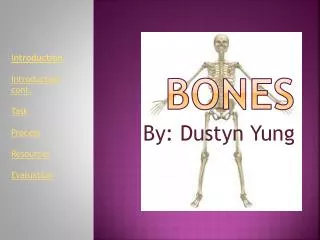
Download
1 / 49
490 likes | 509 Vues
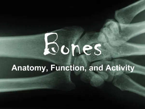
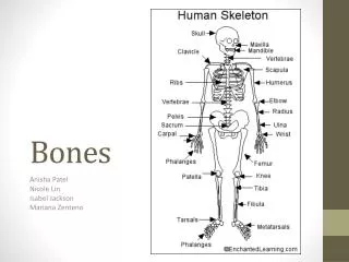
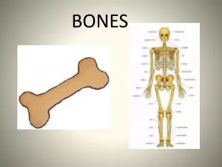
Bones. Bones are rigid substance that constitute the skeletal system There are 206 separate bones in an adult. The largest bone in the human body is the femur and the smallest bones are auditory ossicles. Structural components. Tissues that make up bone are:

E N D
Bones Bones are rigid substance that constitute the skeletal system There are 206 separate bones in an adult. The largest bone in the human body is the femur and the smallest bones are auditory ossicles.
Structural components Tissues that make up bone are: • Mineralized osseous tissue (bone tissue that gives it rigidity) • Marrow • Endosteum • Periosteum • Nerves • Blood vessels • Cartilage
Functions • Protection (bones can serve to protect internal organs, such as the skull protecting the brain or the ribs protecting the heart and lungs) • Structural frame work (bones provide a frame to keep the body supported) • Movement (bones form joints which enhance individual body parts or the whole body tor manipulation in three-dimensional space)
4. Sound transduction (bones in the middle ear) 5. Synthetic - Blood production (the marrow, located within the medullary cavity of long bones and interstices of cancellous bone produces blood cells in a process called hematopoiesis) 6. Mineral storage (bones act as reserves of minerals important for the body, most notably calcium and phosphorus)
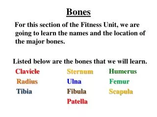
Shape Long bones, Short bones, Flat bones, Irregular bones, Pneumatic bones, Sesamoid bones Embryonic development Membranous bones, Cartilaginous bones, Classification Regionaxial bones & appendicular bone Macroscopic structure Compact bone & spongy bone Microscopic structure Fibrous bone & Lamellar bone
Classification base on shape • Long bones are much longer in length than width. • Characterized by a diaphysis (shaft) and epiphysis at both ends • They are made up mostly of compact bone and less of spongy bone. • It has lesser amounts of marrow, located within the medullary cavity. • Eg: Most bones of the limbs (humerus, femur, radius, ulna,tibia, fibular…
Short bones Are roughly cube-shaped Have only a thin layer of compact bone surrounding a spongy interior. Eg. Carpal bones, tarsal bones …
Flat bones • Are thin and generally curved, with two parallel layers of compact bones sandwiching a layer of spongy bone. • Most of the bones of the skull are flat bones, Scapular, sternum…..
Sesamoid bones Are bones embedded in tendons. They do not possess any periosteum and their ossification also takes place after birth Eg patella…
Irregular bones As implied by the name, their shapes are irregular and complicated Often this irregular shape is due to their many centers of ossification or because they contain bony sinuses. Eg. Vertebra, hip, ethmoid and sphenoid bones
Classification base on embryonic development Membranous bones: The process by which they ossify is called intra-membranous ossification. These bones ossify from mesenchymal condensations in the intrauterine life. Eg: are bones of the skull and facial bones.
Cartilaginous bones: These bones ossify from a cartilage model and this type of ossification is known as intra-cartilaginous ossification. These bones are formed from preformed cartilage models. Eg: bones of limbs, vertebral column and thoracic cage.
Regional Classification Axial bones are bones of the axial skeleton. Eg: are bones of skull and vertebral column Appendicular bone are bones of the appendicular skeleton of the body. Eg: bones of the limbs and girdles.
Classification base on Macroscopic contents Macroscopic approach divides the bones into two categories that are; Compact bone:Occurs when there are more bone tissue and less empty space. Spongy bone: Occurs when there are more empty space and less bone tissue, Eg: Biscuit bone
Classification base on Microscopic contents Microscopic approach divides the bones into following categories; Lamellar bone:The type of bone which are composed of thin plates (lamellae) of bony tissue. Most mature human bones are lamellar bones. Fibrous bone:These have more fibers in them. In humans they are found only in fetus.
A joint is the location at which two or more bones make contact. All of the bones, except the hyoid bone in the neck form a joint. Functions -They are constructed to allow movement -Provide mechanical support -Provide stability -Form a larger unit called skeleton JOINT
Classification of Joints FIBROUS JOINTS Sutures Syndesmoses Gomphoses CARTILAGINOUS JOINTS Primary (Sychondroses) Secondary (Symphyses) SYNOVIAL JOINTS Ball and socket joint Hinge Joints STRUCTURAL CLASSIFICATION Gliding Joints Condyloid Joints Saddle Joints Pivot Joints CLASSIFICATION FUNCTIONAL CLASSIFICATIONSYNARTHROSIS AMPHIARTHROSIS DIARTHROSIS BIOMECHANICAL CLASSIFICATION Uniaxial Biaxial Triaxial
-Structural classification is determined by pattern the bones connect to each other. • Functional classification is determined by the degree of movement between the articulating bones - Bio-mechanical classification is determined by the number of articular surfaces
Structural classificationClasses of joint • Fibrous joint • Cartilaginous joint • Synovial joint
Fibrous joint -Bones are joined by dense irregular connective tissue (collagen fibers) -These type of joints are mostly immovable or may be slightly movable -The fibrous joint is further classified into suture, syndesmose and gomphose.
a. Sutures Join the bones of the skull together to provide protection for the brain and are only. They are immovable joints. b. Syndesmoses Occur in a joint where the bones do not touch each other and are held together by fibrous connective tissue. E.g distal articulation between the tibia and fibula.
c. Gomphoses -Originate from a Greek word gomphos, meaning "bolt" - The only example joints in the human body are the teeth. -The roots of the teeth (the pegs) fit into their (sockets) the alveoli process of the mandible and maxilla -Bundles of collagen fibres pass from the wall of the socket to the root
Cartilaginous joint • Joined by cartilage (fibrocartilage or hyaline) • Cartilaginous joints allow more movement between bones than a fibrous joint but less than the highly mobile synovial joint
Classes of the cartilaginous joint Primary cartilaginous joints “Synchondroses". -Bones are connected by hyaline cartilage -This cartilage ossify with age. -E.g "growth plates" between ossification centers in long bones ( epiphysis and diaphysis) , ribs….. Secondary cartilaginous joints "Symphyses". -Articulating bones covered with hyaline cartilage and have a thick, fairly compressible pad of fibrocartilage between them. . -Usually occurring in the midline. -Eg: manubrosternal joint , intervertebral discs, and the pubic symphysis
Synovial joint • Most common • Most movable type of joint in the body • Capsules (synovial capsule) surrounding the articulating surfaces of bones, within which is lubricating substance (synovial fluid)
Features of synovial joints -Articular surfaces which is covered with hyaline cartilage ( articular cartilage ) reduces friction at the point -Synovial membranelining the joint space (synovial cavity) secretes synovial fluid, which fills the joint space -Synovial fluidprovides lubrication and nourishment to the articular cartilage. -The bones are also attached and held together by strong, tough ligaments made of dense connective tissue (articular capsule ) which prevent dislocation during normal movement.
Types of synovial joints (six ) • Ball and socket joint • Hinge Joints • Gliding Joints • Condyloid Joints • Saddle Joints • Pivot Joints
Ball and socket joint • These joints are formed where the rounded head of one bone fits into the hollow cup-shaped socket of another bone • Ball and socket joints allow for stable movement in several directions (360° ) -Eg: shoulder joint and hip joint.
Hinge Joints -Hinge joints are so named because they resemble hinges find on a door -The articular surfaces are moulded to each other in a manner to permit motion only in one plane (180 ° ) flexion and extension -Very slight degree of rotation or of side-to-side movement - Eg: Interphalangeal joint, elbow joint, knee joint, ankle joint…..
Gliding Joints - Gliding joints allow for smooth movement in several directions along a plane like two plates sliding across each other. • The bones move relative to each other. • Eg: wrist joint
Condyloid Joints -Similar to gliding joints, condyloid joints are somewhat different in that they have an irregular surfaces -This type of joint is like two bowls nested together -Eg: radio-carpal joint of the wrist is an example of a condyloid synovial joint
Saddle Joints • Saddle/ Sellar joint, articulation by the opposing surfaces are reciprocally concave-convex • Saddle joint allows bending motion in several directions without sliding. • Examples is carpal-metacarpal joint of the thumb • permit movement in two planes (3600) a circling motion
Pivot Joints • Pivot joint/Rotary Joint that allows only rotational movement without gliding movement or sideways displacement Examples: - Joint between the first and second cervical vertebrae (atlas-axis) allows for most of the head's range of motion while maintaining the stability of the head on the neck. -Twisting movement of the bones of the forearm (radius and ulna) against the upper arm.
Functional Classification -Synarthrosis permits little or no mobility. Most synarthrosis joints are fibrous joints -Amphiarthrosis permits slight mobility. Most amphiarthrosis joints are cartilaginous joints -Diarthrosis permits a variety of movements. All diarthrosis joints are synovial joints
Bio-mechanical classification Classified according to the number of articulation surfaces UNIAXIAL JOINTS : - Joint motion occurs only in one plane or is said to have one degree of freedom. - Eg: hinge joints, interphalangeal joints…. BIAXIAL JOINTS: - Joint motion occurs in two planes thus there are two degrees of freedom. -Eg: carpometacarpal joint of the thumb, metacarpophalangeal joints of the fingers. TRIAXIAL JOINTS:- Three degrees of motion and can move in three planes. - Eg ball and socket joint