Download
1 / 24
240 likes | 313 Vues
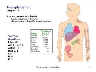
Immunoglobulins Scan pp. 221-228 for background, but you are not responsible for this material. You are responsible for the material on antibodies, pp. 228-233, and the rest of the chapter. Focus on protein-ligand binding

E N D
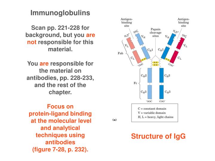
Immunoglobulins Scan pp. 221-228 for background, but you are not responsible for this material. You are responsible for the material on antibodies, pp. 228-233, and the rest of the chapter. Focus on protein-ligand binding at the molecular level and analytical techniques using antibodies (figure 7-28, p. 232). Structure of IgG
Antibodies • - produced and secreted by B lymphocytes • make up 20% of blood protein • each antibody recognizes a specific antigen • An antigen may be a virus, a bacterial cell wall, • a particular protein or other macromolecule • -An individual antibody binds only a particular • molecular structure within the antigen- called the epitope • -Can bind to Fc receptors on macrophages, triggering • phagocytosis of antibody-antigen complex
Host Cell is lysed by virus Soluble antibodies can bind virus and direct them to macrophages to be destroyed
Immunoglobulin G, IgG, the major class of antibody molecule and one of the most abundant proteins in the blood serum. 4 polypeptide chains(2 heavy+2 light chains) Mr 150,000 Antigen-binding fragment Fragment that usually crystallizes
Ribbon model of first complete IgG crystal structure
Binding of IgG to an antigen “Induced fit”: The binding sites of IgG often undergo slight conformational changes as they bind. Common to many protein-ligand interactions.
IgM pentmer- The major antibody in early stages of primary immune response
Phagocytosis of an antibody-bound virus by a macrophage . The Fc regions of the antibodies bind to Fc receptors on the surface of macrophage, triggering it to engulf and destroy virus
ELISA assayImmunoblot Enzyme-linked immunosorbent assay • adsorb sample on polystyrene plate • block empty sites with inert protein • treat with primary antibody, then wash • treat with secondary antibody-enzyme complex, then wash • Add substrate of enzyme contained in secondary antibody • separate sample using gel electrophoresis • transfer proteins electrophoretically to nitrocellulose membrane • block empty sites with inert protein • treat with primary antibody, then wash • treat with secondary antibody-enzyme complex, then wash • add substrate of enzyme contained in secondary antibody
Structure of Skeletal Muscle Z-disk You are responsible for the material on pp. 233-239.
Electron Micrographs of Muscle relaxed contracted
Muscle contraction Relaxed--> Contracted Sliding of thick and thin filaments past each other so that the Z disks in neighboring I bands approach each other
Each thick filament is surrounded by six thin filaments
Myosin Light chains are blue Heavy chains are dark and light pink
Structure of myosin head Light chains Heavy chains