Download
1 / 21
210 likes | 213 Vues
Learn about the Nd:YAG laser capsulotomy procedure, including its benefits, risks, and alternatives. Explore the preparation of the patient before the treatment session and the steps involved in the procedure. Discover the contraindications and potential complications associated with this procedure.

E N D
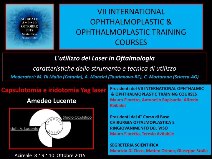
VII INTERNATIONAL OPHTHALMOPLASTIC & OPHTHALMOPLASTIC TRAINING COURSES Presidenti del VII INTERNATIONAL OPHTHALMIC & OPHTHALMOPLASTIC TRAINING COURSES Mauro Fioretto, Antonello Rapisarda, Alfredo Reibaldi Presidenti del 4° Corso di Base CHIRURGIA OFTALMOPLASTICA E RINGIOVANIMENTO DEL VISO Mauro Fioretto, Teresio Avitabile SEGRETERIA SCIENTIFICA Maurizio Di Cicco, Matteo Orione, Giuseppe Scalia Capsulotomia e iridotomia Yag laser Amedeo Lucente L'utilizzo dei Laser in Oftalmologia caratteristiche dello strumento e tecnica di utilizzo Moderatori: M. Di Maita (Catania), A. Mancini (Taurianova-RC), C. Martorana (Sciacca-AG) Acireale 8 · 9 · 10 Ottobre 2015
Disclosure Consulting Free - Carl Zeiss Meditec - Alfa Intes
Nd:YAG è un laser a stato solido che sfrutta come mezzo laser attivo un cristallo di ittrio e alluminio (YAG) drogato al neodimio Nd:Y3Al5O12 • - Nd:YAGNeodymium-dopedYttriumAluminiumGarnet(NY:Y3Al5O12) • 1964 Laser operation of Nd:YAGwas • first demonstrated by J. E. Geusic • Bell Laboratories (New Jersy) • 1980FankhausereAron-Rosa • first YAG capsulotomy • 1064 nm wavelength • Optical breakdown results • inionization, or plasma formation (electromechanical interaction)
Preparation of the patient Before Treatment Session -Complete ophthalmic history and examination - Discussion of proposed procedure, including risks, benefits, and alternatives; signing of informed consent form - Apraclonidine or beta-adrenergic blocking agent - Pupillary dilation (optional) - Determination of visual axis and normal pupillary size: sketch and preliminar laser marker shot - indomethacin drops 0.50% At the Laser - Review of the procedure, the expected pop or click, and the importance of fixation - Application of topical anesthetic if contact lens is to be used - Adjustment of stool, table, chin rest, and footrest for optimal patient comfort - Application of head strap to maintain forehead position - Darkening of the room (optional) - Provision of fixation target for fellow eye - Illumination of target if room is darkened - Photograph the opacity
Sequential capsulotomyphotographs By Roger F. Steinert, MD UCI University of California, Irvine • - Use minimum energy 1 mJif possible • - Identify and cut across tension lines • - Perform a cruciate openinbegin at • 12 o'clock progress toward 6 o'clock • and cut across at 3 and 9 o'clock • - Clean up any residual tags • - Avoid freely floating fragments
Capsulotomy Size • The capsulotomy should be as large as the pupil in isotopic conditions, such as • driving at night, when glare from the exposed capsulotomy edge is most likely • A small opening might be preferred for a patient at high risk of retinal detachment • A small opening in a dense membrane results in excellent optics, analogous to • those of a small pupil • When the capsule is only hazy and transmits images to the retina, a small opening is an improvement but is still suboptimal • As the patient looks up, down, left, and right, the laser can be applied to capsular edges behind the sphincter so that the capsulotomy can be perfectly centered • Capsulotomies may increase in mean area by 32% within 6 weeks with capsular enlargement tending toward sphericity with capsular tag retention • Glare and haze remain a problem for 1‑ and 2‑mm capsular openings, decrease • with a 3-mm opening, and fully resolve only with a 4‑mm capsular opening
Contraindications to laser capsulotomy • Absolute Contraindications • Corneal scars, irregularities, or edema that interfere with target visualization or make optical breakdown unpredictable • Inadequate stability of the eye • - Inadequate stability of the IOL • Relative Contraindications • - Known or suspected cystoid macular edema CME • - Active intraocular inflammation • - High risk for retinal detachment
Complications • IntraocularPressure Elevationgreater than 10 mmHg have been observed in 15% to 67% peaks at 3 to 4 hours, decreases but may remain elevated at 24 hours, and usually returns to baseline at 1week • Cystoid Macular Edema CME0.55% to 2.5% • Retinal detachment 0.08% to 3.6% • Asymptomatic retinal breaks were found at a rate of 2.1% within 1 month • Intraocular Lens Damage, Pitting of IOLs occurs in 15% to 33% of eyes not visually significant, although rarely the damage may cause sufficient glare and image degradation that the damaged IOL must be explanted • Propionibacteriumacnes endophthalmitishas been reported • Iritispersisting for 6 months has been reported in less than 1% • - Macular holes have rarely • Specular microscopic studies have reported corneal endothelial cell loss of 2.3% to 7% • IOL dislocation IOL movement and refractive changes
Conclusions An Overview of Nd:YAG Laser Capsulotomy Eyyup Karahan Duygu Er Suleyman Kaynak Department of Ophthalmology, Izmir, Turkey Review Med Hypothesis Discov Innov Ophthalmol. 2014; 3(2) In conclusion, some complications especially rise in IOP and macular thickness seems to be unavoidable after Nd: YAG laser capsulotomy. Using less total energy and performing smaller capsulotomies are practical choices to decreasecomplications after Nd:YAG capsulotomy
Optical breakdown results in ionization, or plasma formation in the ocular tissue
- Impact point offset by 30 to 200 µm behind the focal plane - Constant pulse duration of 4 nanoseconds - 8/10 µm spot diameter - Minimum energy from 0.5 mJ - Energy adjustable up to 10 mJ
Iridotomy Background Laser peripheral iridotomy (LPI) is the preferred procedure for treating angle-closure glaucoma caused by relative or absolute pupillary block. LPI eliminates pupillary block by allowing the aqueous to pass directly from the posterior chamber into the anterior chamber, bypassing the pupil. LPI can be performed with an argon laser, with a Nd:YAG laser, or, in certain circumstances, with both
Indications • Acute angle-closure glaucoma • Chronic angle-closure glaucoma • Fellow eye of acute angle-closure glaucoma • Narrow/occludable angle • Miscellaneousconditions, includingphacomorphicglaucoma, aqueousmisdirection, nanophthalmos, pigmentarydispersion syndrome, and plateau iris syndrome
Contraindications • Corneal edema • Corneal opacity • Flat anterior chamber
Periprocedural Care • Patient Education/InformedConsent • Nd:YAGlaser an argon laser or both are needed • Usinga contact lens makes the procedureeasier • Abraham lens or a Wise lens • Iridotomybe at least 200/500 μm in size • Gonioscopyis used to assess the anterior chamber angle and AS-OCT • Retroillumination direct and indirect Abrham +66 diopter planoconvex button
Technique • The iridotomy site should be in the peripheral third • A crypt or a thinned area of the iris is recommended • Most ophthalmologists place the iridotomybetween11 o’clock and 1 o’clock, where it is superiorly covered by the lids • Aberrations are lessfrequenta superior site • In patients with blue or green irides • LPI can be performed with a Nd:YAGlaser, using the following • settings: Power - 4-8 mJ, Pulses/burst - 1-3 (the author prefers 2), • Spot size Fixed • - In patients with dark brown irides • First, the argon laser is employed to remove the anterior border of • the iris, using the following settings: Power - 300-400 mW, Spot • size - 50-100 mm, Duration - 0.05 seconds
Complications of Procedure • Postoperativeintraocular pressure spike IOP occurs it is usually in the first hour (as many as 70% of cases) or, less commonly, in the second hour (as many as 40% of cases) • Anterior uveitisis usually mild and can be successfully treated with topical steroids • Iris bleeding and hyphema(50% of patients ) • Corneal decompensation • Closure of the iridotomy site is rare, especially when the Nd:YAG laser is used
Albert Einstein (Ulma, 14 marzo 1879 – Princeton, 18 aprile 1955) “Tutto dovrebbe essere reso il più semplice possibile, ma non più semplicistico”