Download

1 / 16
750 likes | 3.36k Vues
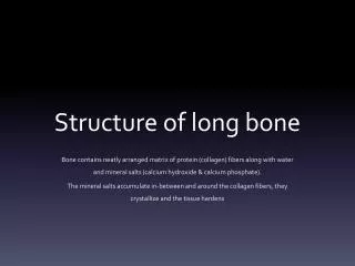
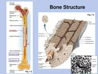
Bone Structure. The diaphysis is the shaft or body of a long bone. The epiphyses form the distal and proximal ends of a long bone. The metaphyses are the areas where the epiphyses and diaphysis join. Bone Structure. In adolescents, through the end of

E N D
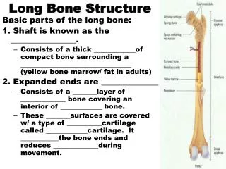
Bone Structure • The diaphysis is the shaft or body of a long bone. • The epiphyses form the distal and proximal ends of a long bone. • The metaphyses are the areas where the epiphyses and diaphysis join.
Bone Structure • In adolescents, through the end of active growth, the epiphysis of the long bones contains hyaline cartilage and forms an “epiphyseal growth plate”. • The growth plate is always actively dividing and causing the bone to elongate from each end.
Bone Structure • In adults, the epiphyseal cartilage is no longer present and elongation of bones has stopped. • The epiphyseal growth plate becomes an “epiphyseal line”, as growing cartilage is replaced by calcified bone. • The epiphyseal line is visible externally and on X-rays.
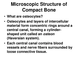
Histology of Bone Tissue • Compact Bone contains units called osteons or Haversian systems formed from concentric lamellae (rings of calcified matrix). • Interstitial lamellae between osteons are left over fragments of older osteons.
Histology of Bone Tissue • Outer circumferential lamellae encircle the bone beneath the periosteum. • Inner circumferential lamellae encircle the medullary cavity.
Histology of Bone Tissue • Lacunae are small spaces between the lamellae which house osteocytes. • Canaliculi are small channels filled with extracellular fluid connecting the lacunae.
Histology of Bone Tissue • Blood and lymphatic vessels are found in the osteon’s Central canal. • Perforating (Volkmann’s) canals allow transit of these vessels to the outer cortex of the bone.
Histology of Bone Tissue • Spongy bone lacks osteons. Instead, lamellae are arranged in a lattice of thin columns called trabeculae. • Trabeculae of spongy bone support and protect the red bone marrow and are oriented along lines of stress (helps bones resist stresses without breaking). • Hematopoiesis (blood cell production) occurs in spongy bone.
Histology of Bone Tissue • Within each trabecula of spongy bone are lacunae . • As in compact bone, lacunae contain osteocytes that nourish the mature bone tissue from the blood circulating through the trabeculae.
Histology of Bone Tissue • The interior of long bones is made up primarily of spongy bone. The use of spongy bone lessens overall bone weight.
Blood and Nerve Supply of Bone • Bone is richly supplied with blood; Periosteal arteries and veins supply the periosteum and compact bone. • Nerves accompany the blood vessels (this is often the case.) • The periosteum is rich in sensory nerves sensitive to tearing or tension (as anyone who has bruised their shin will tell you!)
Bone Formation • Ossification or osteogenesis is the process of forming new bone. Bone formation occurs in four situations: • Formation of bone in an embryo • Growth of bones until adulthood • Remodeling of bone • Repair of fractures
Bone Formation • Osteogenesis occurs by two different methods, beginning about the 6th week of embryonic development. • Intra-membranous ossification produces spongy bone. • This bone may subsequently be remodeled to form compact bone. • Endochondral ossification is a process whereby cartilage is replaced by bone. • Forms both compact and spongy bone.
Bone Formation • Intra-membranous ossification is the simpler of the two methods. • It is used in forming the flat bones of the skull, mandible, and clavicle. • Bone forms from mesenchymal cells that develop within a membrane – without going through a cartilage stage (recall that mesenchyme is the tissue from which almost all other C.T. develop.) • Many ossification centers.