Download
1 / 45
790 likes | 2.01k Vues
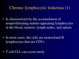
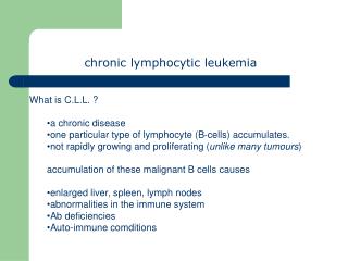
chronic lymphocytic leukemia. is the most common type of leukemia . CLL involves a particular subtype of white blood cells, which is a lymphocyte called a B cell . . The definition of CLL includes >5000 CLL-phenotype B-cell lymphocytes per cubic millimeter.

E N D
is the most common type of leukemia. CLL involves a particular subtype of white blood cells, which is a lymphocyte called a B cell.
The definition of CLL includes >5000 CLL-phenotype B-celllymphocytes per cubic millimeter.
CLL is a disease of adults, but in rare cases it can occur in teenagers and occasionally in children (inherited). Most (>75%) people newly diagnosed with CLL are over the age of 50, and the majority are men.
Although not originally appreciated, CLL is now felt to be identical to a disease called small lymphocytic lymphoma (SLL), a type of non-Hodgkin's lymphoma which presents primarily in the lymph nodes. The World Health Organization considers CLL and SLL to be "one disease at different stages, not two separate entities".
Epidemiology • CLL is a disease of older adults and is rarely encountered in individuals under the age of 40. Thereafter the disease incidence increases with age. • In the United States during 2009, about 16,000 new cases are expected to be diagnosed, and 4,400 patients are expected to die from CLL. Because of the prolonged survival, which was typically about ten years in past decades, but which can extend to a normal life expectancy, the prevalence (number of people living with the disease) is much higher than the incidence (new diagnoses).
Symptoms and signs Mostpeople are diagnosed without symptoms as the result of a routine blood test that returns a high white blood cell count. Uncommonly, CLL presents as enlargement of the lymph nodes without a high white blood cell count or no evidence of the disease in the blood. This is referred to as small lymphocytic lymphoma. Insome individuals the disease comes to light only after the neoplastic cells overwhelm the bone marrow resulting in anemia producing tiredness or weakness.
C / P : (A) Symptoms :- - -> No - - ->may be glandular swelling - - -> loss of weight and general weakness. - - -> fever. Mediastinal--> cough - - -> pressure symptoms : liver --> jaundice
(B) Signs:---> low grade fever --->LN ↑(Usually generalized, moderately enlarged , discrete, firm, not tender( ---> abdomen:- splenomegally: less huge than in CML, liver enlargement may be due to: ---> Leukemic infiltrations ---> skin : nodules, dermatities ---> CNS infiltration ---> laryngeal or salivary gland infiltration
PRESSURE BY LYMPHADEMOPATHY: -Mediastinal syndrome : (cough, hoarsness of voice, dilated veins on the chest wall ,congested non pulsating neck veins, may be +veD'Espine sign, may be homer's syndrome, Dysphagia). - at portahepatis ---> obstructive jaundice
Diagnosis • The disease is easily diagnosed. CLL is usually first suspected by the presence of a lymphocytosis. This frequently is an incidental finding on a routine physician visit. The presence of a lymphocytosis in an elderly individual should raise strong suspicion for CLL and a confirmatory diagnostic test, in particular flow cytometry, should be performed unless clinically unnecessary.
INVESTIGATIONS : Blood Picture : - RBCs: early normal, late : anaemia, may be autoimmune haemolytic anemia. - WBCs:- TC: 60,000 - 200,000 but us.<100.000 - DC:- 90% mature lymphocytes may be few blast cells Platelets : early normal, late ↓ Bone marrow Aspiration : increase number of lymphocytes in the bone marrow (more than1/3 of total population).
The diagnosis of CLL is based on the demonstration of an abnormal population of B lymphocytes in the blood, bone marrow, or tissues that display an unusual but characteristic pattern of surface markers: cluster of differentiation5 (CD5) and cluster of differentiation23 (CD23). In addition, all the CLL cells within one individual are clonal, that is genetically identical. In practice, this is inferred by the detection of only one of the mutually exclusive antibody light chains, kappa or lambda, on the entire population of the abnormal B cells
The combination of the microscopic examination of the peripheral blood and analysis of the lymphocytes by flow cytometry to confirm clonality and marker molecule expression is needed to establish the diagnosis of CLL.
Morphologically, the cells resemblenormal lymphocytes under the microscope, although slightly smaller, and are fragile when smeared onto a glass slide giving rise to many broken cells (smudge cells).
Differential diagnosis Hematologic disorders that may resemble CLL in their clinical presentation, behavior, and microscopic appearance include: mantle cell lymphoma, marginal zone lymphoma, B cell prolymphocytic leukemia, and lymphoplasmacytic lymphoma.
All the B cell malignancies of the blood and bone marrow can be differentiated from one another by the combination of cellular microscopic morphology, marker molecule expression, and specific tumor-associated gene defects.
A flow cytometer is necessary for cell marker analysis and the detection of genetic problems in the cells may require visualizing the DNA changes with fluorescent probes by fluorescent in situ hybridization (FISH).
Monoclonal B-cell lymphocytosis (MBL) • indicate a monoclonal B cell population in a person with less than 5000 B lymphocytes per milliliter (or 5.0 x 10⁹ B lymphocytes/L), no enlarged lymph nodes or enlarged liver and/or spleen or other indications of a lymphoproliferative disorder. • MBL has been found in less than 1% of asymptomatic adults under age 40, and in around 5% of adults older than 60
Recent studies suggest that CLL is very often preceded by MBL, and that MBL progresses to CLL requiring treatment at a rate of around 1-2% per year. Advancing age and high initial B cell count predispose to progression from MBL to CLL; however, only a small fraction of people with MBL die because of CLL.
Thus, MBL could be regarded as a premalignant condition from which some cases progress to CLL .No treatment is required, but follow-up might be able to detect new diagnoses of CLL.
B cell prolymphocytic leukemia (B PLL), is a related but more aggressive disorder, has cells with similar phenotype but that are significantly larger than normal lymphocytes and have a prominent nucleolus. The distinction is important as the prognosis and therapy differs from CLL.
Hairy cell leukemia is also a neoplasm of B lymphocytes but the neoplastic cells have a distinct morphology under the microscope (hairy cell leukemia cells have delicate, hair-like projections on their surface) and unique marker molecule expression.
Clinical staging Staging, determining the extent of the disease, is done with the Rai staging system or the Binet classification and is based primarily on the presence, or not, of a low platelet or red cell count. Early stage disease does not need to be treated.
Staging is useful in chronic lymphocytic leukemia (CLL) to predict prognosis and also to stratify patients to achieve comparisons for interpreting specific treatment results. Anemia and thrombocytopenia are the major adverse prognostic variables.
Rai Staging System Stage 0 • Stage 0 CLL is characterized by absolute lymphocytosis (>15,000/mm3) only • Stage I • Stage I CLL is characterized by absolute lymphocytosis with lymphadenopathy without hepatosplenomegaly, anemia, or thrombocytopenia. • Stage II • Stage II CLL is characterized by absolute lymphocytosis with either hepatomegaly or splenomegaly with or without lymphadenopathy. • Stage III • Stage III CLL is characterized by absolute lymphocytosis and anemia (hemoglobin <11 g/dL) with or without lymphadenopathy, hepatomegaly, or splenomegaly. • Stage IV • Stage IV CLL is characterized by absolute lymphocytosis and thrombocytopenia (<100,000/mm3) with or without lymphadenopathy, hepatomegaly, splenomegaly, or anemia. • Binet Classification
Binet Classification • Clinical stage A* • Clinical stage A CLL is characterized by no anemia or thrombocytopenia and fewer than three areas of lymphoid involvement (Rai stages 0, I, and II). • Clinical stage B* • Clinical stage B CLL is characterized by no anemia or thrombocytopenia with three or more areas of lymphoid involvement (Rai stages I and II). • Clinical stage C • Clinical stage C CLL is characterized by anemia and/or thrombocytopenia regardless of the number of areas of lymphoid enlargement (Rai stages III and IV).
the presence of either cluster of differentiation38 (CD38) or Z-chain–associated protein kinase-70 (ZAP-70) may be surrogate markers of high risk subtype of CLL. Their expression correlates with a more immature cellular state and a more rapid disease course.
Fluorescence in situ hybridization (FISH) Four main genetic aberrations are recognized in CLL cells that have a major impact on disease behavior. • Deletions of part of the short arm of chromosome 17 (del 17p) . Patients with this abnormality have significantly short interval before they require therapy and a shorter survival. This abnormality is found in 5-10% of patients with CLL. • Deletions of the long arm on chromosome 11 (del 11q) are also unfavorable although not to the degree seen with del 17p. This abnormality occurs infrequently in CLL (5-10%).
Fluorescence in situ hybridization (FISH) • Trisomy 12, an additional chromosome 12, is a relatively frequent finding occurring in 20-25% of patients and imparts an intermediate prognosis. • Deletion of the long arm of chromosome 13 (del 13q) is the most common abnormality in CLL with roughly 50% of patients with cells containing this defect. These patients have the best prognosis and most will live many years, even decades, without the need for therapy.
Array-based Karyotyping Array-based karyotyping is a cost-effective alternative to FISH for detecting chromosomal abnormalities in CLL.
Virtual Karyotype Virtual Karyotype (also Array comparative genomic hybridization, CMA, Chromosomal Microarray Analysis, Microarray-based comparative genomic hybridization, array CGH, a-CGH, aCGH, or molecular karyotyping. If using SNP-based arrays, also SNP array karyotyping, molecular allelokaryotyping or SOMA) detects genomic copy number variations at a higher resolution level than conventional karyotyping or chromosome-based comparative genomic hybridization (CGH).
Treatment CLL treatment focuses on controlling the disease and its symptoms rather than on an outright cure. CLL is treated by chemotherapy, radiation therapy, biological therapy, or bone marrow transplantation. Symptoms are sometimes treated surgically (splenectomy removal of enlarged spleen) or by radiation therapy ("de-bulking" swollen lymph nodes).
Decision to treat While generally considered incurable, CLL progresses slowly in most cases. Because of its slow onset, early-stage CLL is generally not treated since it is believed that early CLL intervention does not improve survival time or quality of life. Instead, the condition is monitored over time to detect any change in the disease pattern.
The decision to start CLL treatment is taken when the patient's clinical symptoms or blood counts indicate that the disease has progressed to a point where it may affect the patient's quality of life. Clinical "staging systems" such as the Rai 4-stage system and the Binet classification can help to determine when and how to treat the patient
Determining when to start treatment and by what means is often difficult; studies have shown there is no survival advantage to treating the disease too early.
Purine analogues Although the purine analogue fludarabine was shown to give superior response rates than chlorambucil as primary therapy,thereis no evidence that early use of fludarabine improves overall survival, and some clinicians prefer to reserve fludarabine for relapsed disease.
Monoclonal antibodies • Monoclonal antibodies are alemtuzumab (directed against CD52) and rituximab (directed against CD20).
Combination chemotherapy Combination chemotherapy options are effective in both newly-diagnosed and relapsed CLL. Recently, randomized trials have shown that combinations of purine analogues (fludarabine) with alkylating agents (cyclophosphamide) produce higher response rates and a longer progression-free survival than single agents: • FC (fludarabine with cyclophosphamide) • FR (fludarabine with rituximab) • FCR (fludarabine, cyclophosphamide, and rituximab) • CHOP (cyclophosphamide, doxorubicin, vincristine and prednisolone)
Stem cell transplantation Allogeneicbone marrow (stem cell) transplantation is rarely used as a first-line treatment for CLL due to its risk. Autologous stem cell transplantation, a lower-risk form of treatment using the patient's own blood cells, is not curative.
Refractory CLL Refractory" CLL is a disease that no longer responds favorably to treatment. In this case more aggressive therapies, including lenalidomide, flavopiridol, and bone marrow (stem cell) transplantation, are considered.Themonoclonal antibody, alemtuzumab (directed against CD52), may be used in patients with refractory, bone marrow-based disease.
COMPLICATIONS (1): • -1/3 of patient ----> auto immune haemolytic anemia • -1/3 of patient ---->Hypogammaglobulinemia • -Average life expectancy ---> 3 - 4 years • -Most patients respond well to chemotherapy and radiotherapy --> long periods of remissions. • -early the patient may remain alive, even without treatment for several years. • Causes of death : • * may be from other causes • * pneumonia • *overwelming infections • *Haemorrhage
Complications(2) Chronic lymphocytic leukemia may transform into Richter's syndrome, a term used to describe the development of high-grade non-Hodgkin lymphoma, prolymphocytic leukemia, Hodgkin disease, or acute leukemia in a patient who has chronic lymphocytic leukemia. Its incidence is estimated to be around 5%.