Download
1 / 75
770 likes | 1.07k Vues
Tissue Repair. Somatic Cell Division Interphase – 95% of cell cycle Organelle duplication, DNA replication, Growth. G1 Phase Metabolically active Organelle duplication, but no DNA replication Duration variable – short in embryonic and cancer cells Prepares for S phase

E N D
Somatic Cell Division Interphase – 95% of cell cycle Organelle duplication, DNA replication, Growth
G1 Phase Metabolically active Organelle duplication, but no DNA replication Duration variable – short in embryonic and cancer cells Prepares for S phase Cells that remain in G1 for a long time = G0 (permanent tissues, such as neural tissue)
S Phase Committed to cell division once this starts DNA and centrosome replication Semi-conservative replication of DNA: two identical daughter genomes
G2 Phase Growth continues Enzymes and proteins synthesized for cell division Determining Cell Stage Cells at different stages of the cell cycle can also be distinguished by their DNA content
Mitotic (M) Phase mitosis plus cytokinesis Mitosis: Prophase Metaphase Anaphase Telophase
Regulation of the Cell Cycle Cell Cycle Lengths Vary by cell type: Embryonic cells 1-Stem cells (e.g., blood cells and epithelial cells) 2-Sperm cells G1 prolonged in stable or permanent cells (called G0) G1 rapid or non-existent in rapidly-dividing cells
Embryonic cells Cell growth not part of cell cycle All energy goes into DNA synthesis So G1 lacking and G2 quite short Each round of division subdivides original cytoplasm into smaller and smaller cells, Until adult cell size is reached
Cell-Cycle Checkpoints G1 checkpoint In yeast, called start In animal cells, called restriction point G2 checkpoint Located at boundary between G2 and M phase Proper completion of DNA synthesis required before cell can initiate mitosis Spindle Assembly Checkpoint Boundary between metaphase and anaphase All chromosomes must be properly attached to the spindle
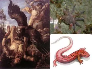
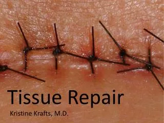
Regeneration refers to growth of cells and tissues to replace lost structures, such as the growth of an amputated limb in amphibians. In mammals, whole organs and complex tissues rarely regenerate after healing
Healing is usually a tissue response to a wound (commonly in the skin), to inflammatory processes in internal organs, or to cell necrosis in organs incapable of regeneration
Regeneration requires an intact connective tissue scaffold. • By contrast, healing with scar formation occurs if the extracellular matrix (ECM) framework is damaged, causing alterations of the tissue architecture.
Repair processes are critical for the maintenance of normal structure and function and survival of the organism. • The healing of skin wounds is just the most common example of repair processes. However, in healthy tissues, repair, in the form of regeneration or healing, occurs after practically any insult that causes tissue destruction.
Epidermal Growth Factor (EGF) and Transforming Growth Factor-α (TGF-α). • Hepatocyte Growth Factor (HGF)/scatter factor • Vascular Endothelial Growth Factor (VEGF). • Platelet-Derived Growth Factor (PDGF). • Fibroblast Growth Factor (FGF • Wound repair: FGFs participate in macrophage, fibroblast, and endothelial cell migration in damaged tissues and migration of epithelium to form new epidermis • TGF-β and Related Growth Hematopoiesis:FGFs have been implicated in the differentiation of specific lineages of blood cells and development of bone marrow stroma. • Transforming Growth Factors. Effects of TGF-β on mesenchymal cells it generally stimulates the proliferation of fibroblasts and smooth muscle cells. TGF-β is a potent fibrogenic agent that stimulates fibroblast chemotaxis, enhances the production of collagen, fibronectin, and proteoglycans. It inhibits collagen degradation by decreasing matrix proteases and increasing protease inhibitor activities. TGF-β is involved in the development of fibrosis in a variety of chronic inflammatory conditions particularly in the lungs, kidney, and liver. TGF-β has a strong anti-inflammatory effect.
The replication of cells is generally stimulated by growth factors or by signaling from ECM components through integrins • To enter the cycle, quiescent cells first must go through the transition from G0 to G1, the first decision step, which functions as a gateway to the cell cycle • Checkpoint activation delays the cell cycle and triggers DNA repair mechanisms
The goal of the repair process is to restore the tissue to its original state. The inflammatory reaction set in motion by the injury contains the damage, eliminates the damaging stimulus, removes injured tissue • Some tissues can be completely reconstituted after injury, such as the repair of bone after a fracture or the regeneration of the surface epithelium in a cutaneous wound. For tissues that are incapable of regeneration, repair is accomplished by connective tissue deposition, producing a scar. This term is most often used in connection to wound healing in the skin, but it is also used to describe the replacement of parenchymal cells by connective tissue, as in the heart after myocardial infarction. • If damage persists, inflammation becomes chronic, and tissue damage and repair may occur concurrently. Connective tissue deposition in these conditions is usually referred to as fibrosis.
Repair begins early in inflammation. • Fibroblasts and vascular endothelial cells begin proliferating to form a specialized type of tissue that is the hallmark of healing, granulation tissue. • Pink, soft, granular appearance on the surface of wounds, but it is the histologic features that are characteristic: • The formation of new small blood vessels (angiogenesis) and the proliferation of fibroblasts • New granulation tissue is often edematous.
Angiogenesis from Pre-Existing Vessels • Angiogenesis from Endothelial Precursor Cells • Growth Factors and Receptors Involved in Angiogenesis
Vasodilation in response to nitric oxide and VEGF-induced increased permeability of the pre-existing vessel • Proteolytic degradation of the BM of the parent vessel by metalloproteinases and disruption of cell-to-cell contact between endothelial cells of the vessel by plasminogen activator • Migration of endothelial cells toward the angiogenic stimulus • Proliferation of endothelial cells, just behind the leading front of migrating cells • Maturation of endothelial cells, which includes inhibition of growth and remodeling into capillary tubes • Recruitment of periendothelial cells (including pericytes for small capillaries and vascular smooth muscle cells for larger vessels) to support the endothelial tubes and form the mature vessel.
SCAR FORMATION Growth factors and cytokines released at the site of injury induce fibroblast proliferation and migration into the granulation tissue framework of new blood vessels and loose ECM that initially forms at the repair site. • (1) emigration and proliferation of fibroblasts in the site of injury • (2) deposition of ECM, and (3) tissue remodeling. • Fibroblast Migration and Proliferation • Migration of fibroblasts to the site of injury and their subsequent proliferation are triggered by multiple growth factors, including TGF-β, PDGF, EGF, FGF, and the cytokines IL-1 and TNF • Macrophages are important cellular constituents of granulation tissue, clearing extracellular debris, fibrin, and other foreign material at the site of repair. • Fibroblast migration and proliferation, increased synthesis of collagen and fibronectin, and decreased degradation of ECM by metalloproteinases TGF-β is also chemotactic for monocytes and causes angiogenesis in vivo, possibly by inducing macrophage influx. • ECM Deposition and Scar Formation : As repair continues, the number of proliferating endothelial cells and fibroblasts decreases. • (PDGF, FGF, TGF-β) and cytokines (IL-1, IL-13) • Net collagen accumulation, however, depends not only on increased collagen synthesis but also on decreased degradation. • The replacement of granulation tissue with a scar involves transitions in the composition of the ECM. • The balance between ECM synthesis and degradation results in remodeling of the connective tissue framework-an important feature of both chronic inflammation and wound repair.
Components of the ECM • Collagens for tensile strength. • Elastin important in large vessels, uterus, skin and ligament • Proteoglycans and hyaluronan forms hydrated gels. Proteoglycans also store bFGF • Adhesive Proteoglycans connects ECM-ECM and ECM-cell integrins . The ECM -cell interaction can activate the same pathways as growth factors
The surgeon told his patient that woke up after having been operated: "I'm afraid we're going to have to operate you again. Because, you see, I forgot my rubber gloves inside you.""Well, if it's just because of them, I'd rather pay for them if you just leave me alone."
1) Which of the following is associated with acute inflammation? • a) Neutophils • b) Macrophages • c) Lymphocytes • d) Tissue fibrosis • e) Tissue necrosis