Download
1 / 20
200 likes | 352 Vues
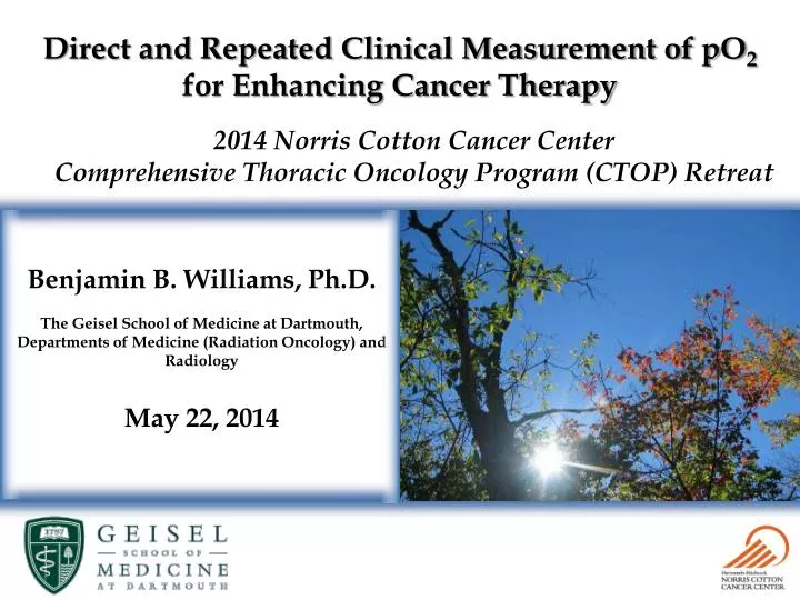
Direct and Repeated Clinical Measurement of pO 2 for Enhancing Cancer Therapy. 2014 Norris Cotton Cancer Center Comprehensive Thoracic Oncology Program (CTOP) Retreat . Benjamin B. Williams, Ph.D.

E N D
Direct and Repeated Clinical Measurement of pO2 for Enhancing Cancer Therapy 2014 Norris Cotton Cancer Center Comprehensive Thoracic Oncology Program (CTOP) Retreat Benjamin B. Williams, Ph.D. The Geisel School of Medicine at Dartmouth, Departments of Medicine (Radiation Oncology) and Radiology May 22, 2014
EPR: Background Fundamentals • EPR is a form of magnetic resonance spectroscopy that measures the absorption of RF energy by unpaired electrons in a magnetic field. • The physics is similar to that of MRI, but unpaired -e are detected. • Features and considerations: • Non-ionizing modality • Spectroscopic data available • Sensitive to the magnetic environment • In most cases exogenous spin probes must be introduced. • Lower magnetic fields • Higher RF frequencies, shallower penetration • Rapid relaxation of electron spins affects detection strategies Clinical EPR spectrometer Subject positioned for oximetry using India ink as reporter
Unique or Exceptional Capabilities of In Vivo EPR In vivo EPR is sensitive to a wide array of physiologic parameters. This information is observed through simple changes in the shapes of the detected spectra. • Partial pressure of oxygen (pO2; using oxygen-sensitive paramagnetic materials) • Free radicals observed directly or by spin trapping, including oxygen-, carbon-, and sulfur-centered radicals • Nitric Oxide (spin trapping) • Redox status (using metabolism of nitroxides) • Thiol groups (using specific nitroxides) • pH (using specific nitroxides) • Perfusion (using washout of paramagnetic tracers) • Metal ions (in paramagnetic states such as chromium, manganese) • Absorbed dose of radiation (in teeth, nails, or bones)
EPR Sensitivities : Oximetry Collisions between the spin probe and molecular O2 promote relaxation and broaden the linewidth of the observed spectrum. LiPc
Significance of Hypoxia for Cancer Therapy • Hypoxia plays crucial roles in tumor development and treatment. • The response of tumors to cytotoxic therapies, especially ionizing radiation, is critically dependent on pO2. • Hypoxia may promote metastatic growth and tumor phenotypes that are more resistant to therapy. • Hypoxia is dynamic: diffusion, perfusion (incl. cycling), anemic Tatum JL, Kelloff GJ, Gillies RJ, et al. Hypoxia: Importance in tumor Biology, noninvasive measurement by imaging, and value of its measurement in the management of cancer therapy. IntJ Radiat Biol. 2006 Oct;82(10):699-757.
Significance of Hypoxia for Cancer Therapy • Hypoxia plays crucial roles in tumor development and treatment. • The response of tumors to cytotoxic therapies, especially ionizing radiation, is critically dependent on pO2. • Hypoxia may promote metastatic growth and tumor phenotypes that are more resistant to therapy. • Hypoxia is dynamic: diffusion, perfusion (incl. cycling), anemic Hockel M, Schelnger K, Aral B, Mitze M, Schaffer U, Vaupel P. Association between Tumor Hypoxia and malignant Progression in Advanced Cancer of the Uterine Cervix. Can Res. 1996 Oct;56:4509-4515. Eric J. Hall, Amato J. Giaccia. Radiobiology for the radiologist.Lippincott Williams & Wilkins, 2006; 546p.
Optimize Delivery Timing of dose Application of pO2modifiers Combination therapies * * Points of pO2 measurement * * Design Select patients Tumor profiling pO2 heterogeneity Assessment pO2 redistribution Vascular remodeling Monitor normal tissues Overall goal • To establish EPR oximetry as a clinical tool for noninvasive monitoring of tumor oxygen in human subjects, under conditions that are fully compatible with clinical practice Treatment Outcome Improved Survival Initiation of intervention Therapeutic Cycle
Capabilities of EPR Oximetry • EPR oximetry can provide repeated and direct measurements of absolute pO2 in tumors and other tissues. • Following introduction of the reporters (injection or implantation), the measurements are non-invasive • Measurements are made in the tissue at the site of interest • Measurements can be made continuously or repeatedly as needed, often indefinitely, at the same site • Sub-mm resolution can be achieved • The measurements are especially sensitive and accurate at the low levels of pO2 (sensitivity to D < 1 mmHg) • Compatibility with complementary techniquesfor imaging and measurement • Immediate clinical applicabilityusing India ink, advanced techniquesin stages of regulatory approval
EPR Oximetry with India Ink • Patient preparation - Injection of India ink • India ink formulated with Printex-U carbon black (200mg/ml) in 0.9% NaCl and 1.25% CMC • Sterilized via autoclave prior to injection • 20-50mL injected into tissue of interest using 22 gauge needle • 1-10mm depth • Measurements • Clinical whole-body spectrometer with temperature control • EPR data collection (10s/scan, 1-30 min/set) • Measurements repeated over the course of treatment as desired Ink injection and calibrated pO2 response
Preliminary Measurements in Humans • Tumor pO2 has been measured in 14 volunteers. • Various tumor types of superficial tumors • Measurement sites have ranged from the feet to the scalp. • Tumor pO2 varied among the patients studied and over the course of treatment. • Tumors were tested for response to changes of inhaled oxygen gas. • Large differences among tumors were found showing potentially very important implications for improving personalized therapy based on the oxygen levels in that person’s tumor.
Tumor pO2 was monitored in melanoma metastases at two sites, in the scalp and neck, during the course of radiation treatment (66 Gy). Spectra were recorded immediately before and after each fraction while the patient inspired room air. These results indicate hypoxic environments that vary on a day-to-day basis, but show little acute response in pO2 due to radiation. pO2 Variability across Serial Measurements
Oximetry with Implantable Resonators The coupling loop of the implantable resonator is placed subcutaneously and the detection loop(s) are inserted into the tissue of interest. • The development of implantable resonators will extend applicability of EPR oximetry • Implantable resonators extend the range of EPR oximetry to deeper tissues. • ‒ Improved SNR at all depths • ‒ Improved spatial localization • Ability to use optimal materials for added sensitivity • ‒ Biocompatible coating contains paramagnetic oximetric material • ‒ Ability to remove implants following use The implantable resonators can be fabricated with great flexibility in configuration. Installation for deep tissue measurements of pO2 in the flank of a pig
pO2 Assessment following Sub-lobar Resection with Adjuvant SABR • NCCC-ACS-IRG funded effort – PI: Philip Schaner (Rad. Onc), Co-I: Erkmen, Williams, Hou, Black • Proposes that local control of early-stage NSCLC following sub-lobar resection could be improved with adjuvant SABR • Efficacy of radiation is related to tissue pO2, which may be diminished following surgical disruption. • Tissue pO2 will allow a more complete understanding of the potential of adjuvant SABR in the post-operative setting. • Primary Objective: to evaluate the technical feasibility of adjuvant stereotactic ablative radiotherapy (SABR) after sub-lobar resection, and obtain preliminary safety data, in a large animal model (pig) • Secondary Objective: to evaluate oxygenation using a novel application of EPR in the pulmonary parenchyma, both after sub-lobar resection and post-operative SABR. • Status: Entering in vivo stage, following procedural evaluation and refinements with cadaveric animals
PPG Layout: Projects & Cores PROJECT 1 PROJECT 1:Oxygen measurements in human tumors using EPR oximetry with carbon-based sensors (PI: Gallez) PROJECT 2: Monitoring of pO2 in human tumors using EPR oximetry with implantable oxygen-sensing probe, OxyChip (PI: Kuppusamy) PROJECT 3: Oxygen measurements in deep-seated human tumors using EPR oximetry with implantable deep-tissue oxygen sensors (PI: Swartz) CORE A: Administrative Core CORE B: Instrument/Resource Core CORE C: Biostatistics Core Carbon CORES A,B,C Primary: India ink OxyChip ImOS PROJECT 2 PROJECT 3 Deep-tissue Sensor OxyChip Primary: India ink OxyChip ImOS
Synopsis of Project 1 Oxygen measurements in human tumors using EPR oximetry with carbon-based sensors (PI: Gallez; Sites: Dartmouth, Brussels, Emory, UPenn) • Make use of already approved materials (India ink in USA and charcoal in Europe) for human measurements • pO2 measurements in superficial tumors, up to 10 mm • depth • Dartmouth has performed initial clinical measurements • in human tumors • Studies will be performed to establish: • Intra-tumor and inter-tumor variations of pO2 levels using repeated measurements over a period of weeks in the same tumor • Tumor response to hyperoxygenation treatments • Comparison of EPR pO2 data with those obtained using indirect methods such as PET, MRI, and hypoxia gene signatures • Monitor pO2 dynamics in tumors and normal tissue following radiation therapy • Role of oxygen in radiation-induced fibrosis • Tumors: Head & Neck (oral), Cervical, Breast, Mycosis Fungoides
Synopsis of Project 2 Monitoring of pO2 in human tumors using EPR oximetry with an implantable oxygen-sensing probe (OxyChip) (PI: Kuppusamy; Sites: Dartmouth, Emory) • Will use OxyChip – a newly developed, high-sensitive, PDMS-coated implantable/retrievable sensor for EPR oximetry • Awaiting FDA approval for Investigational Device Exemption • The sensor has been tested and validated in preclinical animal models • Repeated pO2measurements will be made in superficial tumors (up to 10-mm depth) • Studies will be performed to establish: • Safety and efficacy for repeated measurements of pO2 in human subjects • Monitor temporal changes in tumor pO2 over a period of weeks to months • Tumor response to hyperoxygenation treatments • Comparison of EPR pO2 data with hypoxia gene signatures • Monitor pO2 dynamics in tumor and normal tissue following radiation therapy • Role of oxygen in neo-adjuvant trastuzumab therapy for HER2+ breast cancer • Tumors: Head & Neck (oral), Cervical, Breast (HER2+), Mycosis Fungoides
Synopsis of Project 3 Oxygen measurements in human tumors using EPR oximetry with implantable deep-tissue oxygen sensors (PI: Swartz; Sites: Dartmouth, Emory) • Will use implantable deep-tissue oxygen sensors for repeated measurement of pO2 in deep-sited tumors • Seek FDA approval for Investigational Device Exemption • The sensor has been tested and validated in animal models • pO2 measurements will be made in tumors deeper than 1 cm • Studies will be performed to establish: • Safety and efficacy for repeated measurements of pO2 in human subjects • Monitor changes in tumor pO2 over a period of weeks to months • Tumor response to hyperoxygenation treatments • Comparison of EPR pO2 data with hypoxia gene signatures • Monitor pO2 dynamics during concurrent treatment with radiation and oxygen-modifying interventions • Tumors: Head & Neck (lymph node), high-grade sarcoma, glioblastoma
Clinical EPR Oximetry Summary • In vivo EPR oximetry has been used successfully in the clinical setting to make repeated non-invasive direct measurements of tissue pO2. • pO2 in peripheral tissues can be measured, including responses to changes in FIO2 and perfusion • In most tumors, baseline pO2 values were observed to be quite low (>10mmHg) and the application of inhaled oxygen led to dramatic increases in tumor pO2. • Tumor pO2 values have varied among the patients studied and over the courses of treatment • Different responses of tumor pO2 to increased fractions of inhaled oxygen are observed. • Implantable probes and resonators and are being developed to extend the clinical applicability of EPR oximetry to include deep tumor sites. • Based on the measurements to date, we believe that it is feasible that in vivo EPR oximetry could be used to monitor tumor pO2 in the clinical setting and guide the optimal application of therapies.
Acknowledgements • NIH, Norris Cotton Cancer Center, ACS Institutional Research Grants, • Dartmouth Center for Clinical and Translational Science (DCCTS) • Department of Radiology, Section of Radiation Oncology Investigators Harold SwartzLesley Jarvis Periannan Kuppusamy Phil Schaner Nadeem Khan Eunice Chen Huagang Hou Alan Eastman Ann Flood Eugene Demidenko Bernard Gallez (UCL, Brussels, Belguim)