Download
1 / 19
190 likes | 272 Vues
Clinton Jung Cjung3@jhu.edu Advisor: Bir Bhanu Center for Research in Intelligent Systems August 20, 2009. Understanding and Quantifying the Dancing Behavior of stem cells before attachment. Overview. Background Project Objectives Technical Methods Otsu’s Method

E N D
Clinton Jung Cjung3@jhu.edu Advisor: BirBhanu Center for Research in Intelligent Systems August 20, 2009 Understanding and Quantifying the Dancing Behavior of stem cells before attachment
Overview • Background • Project Objectives • Technical Methods • Otsu’s Method • Connected Components Algorithm • Results and Analysis • Conclusions
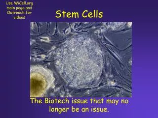
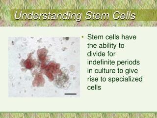
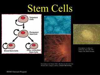
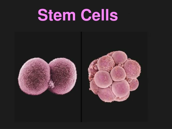
Background • What are stem cells? • Two lines • Embryonic stem cells • Adult stem cells • Five states • Attached to substrate • Unattached • Dancing, or pre-attachment behavior • Death, or apoptosis • Mitosis
Background cont. • Stem cell culture conditions • 37 degrees Celsius • Treated with cigarette smoke—traditional and harm reduction • Mouse embryonic stem cells are used because they are easier to obtain and manipulate • Matragel substrate • Video Capture • Uses Biostation Hardware • Time-lapse style • 3.5 hours, 106 still images
Background cont. • Stem cells alternate between five states • Dancing always comes before a cell attaches • Mitosis only occurs when cells are unattached • Apoptosis is often confused with dancing behavior • Cell Arrangements • Single Cell • Colony
Project Objectives • Crop images for the cells undergoing dancing and play as video for each cell • Develop image segmentation techniques • Find connected components and compute features • Quantify the dancing phenomenon and change in shape
Video Cropped for Dancing Dancing Cells
Observations • Cells undergoing pre-attachment behavior (dancing) • Often have several legs or appendages when dancing. These bulbs are roughly one third or less of the original size of the cell before dancing • A cell may undergo several cycles of detachment, dancing, attachment • May affect the state of surrounding cells and influence them in some manner • Dancing often occurs after mitosis but not necessarily • Cells attached to substrate • Can be identified by an increase in surface area and exhibit a darker inner intensity value • When cells attach, they lose their circular shape and instead become noncircular. There were cells that were semi-attached
Technical Methods • Image Segmentation- Process of dividing an image into different segments (sets of pixels). This technique can be used to locate objects and boundaries based on lines, curves, contrasts • Benefits • Automatically reduce image to simpler one to analyze • Identify different components and features of an image • Simpler, processed images can then be analyzed
Otsu’s Method • Step 1: Input Original Image and convert to grayscale • Step 2: Find threshold automatically using histogram. Two groups of pixels created such that the intra-class variance is minimal and inter-class variance maximal • Step 3: Split image into two classes (binary) based on the threshold value. This final image will have pixel values of either 0 or 255. Number of Pixels Grayscale Value Calculated Threshold Value: 138
Connected Components Algorithm • Step 1: Input Original Image which must be a binary image • Step 2: For each pixel, examine surrounding 8 pixels • If the pixel is neighboring, assign label 1 • If pixel is not neighboring, assign label 0 • Step 3: Continue to check each pixel line by line until entire image is checked, resulting in a matrix of 1’s and 0’s • Step 4: Convert image back to rgb in order to display components in colors.
Analysis • Expand connected components program to count the number of pixels in a cell undergoing pre-attachment behavior and plot this value over time Total Number of Pixels vs. Time Frame 85 Total Number of Pixels in connected component frame Frame 41 Time (Video Frame Number)
Analysis cont. • Expand program to compute average grayscale value of a dancing cell over time and examine the averages and standard deviations of these values over time per frame. Average Grayscale Value vs. Time Average Grayscale Value Time (Video Frame Number)
Analysis cont. Grayscale Standard Deviation vs. Time Grayscale Standard Deviation Time (Video Frame Number)
Conclusions • A preliminary calculation of the changes in pixel count for a cell undergoing pre-attachment behavior displays no periodic behavior. Pixel count data is also consistent with our hypothesis of a high pixel count for cells in dancing and a low count for a cell in attachment. • The graphical analysis shows that the cells in the dancing, attached, and unattached states have very distinct average grayscale values per frame over the course of the video and can potentially be automatically differentiated using this property • However, the standard deviations of these values for the different stem cell states have a more inconsistent pattern which results in interference with each other • The average and standard deviation values can be combined using pattern recognition techniques
Acknowledgements • I would like to extend much gratitude to • Dr. Bir Bhanu for his guidance and advice • Benjamin Guan for his willingness to teach • Jun Wang for the opportunity this summer to do research • Talbot Lab—Dr. Prue Talbot and Sabrina Lin for growing and filming the stem cells • Shubham Debna and Lindsay Kulkin for their collaboration • Other members of the BRITE program and C.R.I.S.