Download
1 / 3
30 likes | 125 Vues
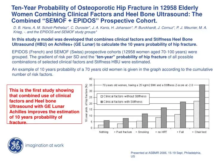
This is the first study showing that combined use of clinical factors and Heel bone Ultrasound with GE Lunar Achilles improves the estimation of 10 years probability of fracture. .

E N D
This is the first study showing that combined use of clinical factors and Heel bone Ultrasound with GE Lunar Achilles improves the estimation of 10 years probability of fracture. Ten-Year Probability of Osteoporotic Hip Fracture in 12958 ElderlyWomen Combining Clinical Factors and Heel Bone Ultrasound: TheCombined “SEMOF + EPIDOS” Prospective Cohort. In this study amodel was developed that combines clinical factors and Stiffness Heel Bone Ultrasound (HBU) on Achilles+ (GE Lunar) to calculate the 10 years probability of hip fracture. EPIDOS (French) and SEMOF (Swiss) prospective cohorts (12958 women aged 70-100 years) were grouped. The gradient of risk per SD and the “ten-year” probability of hip fracture of all possible combinations of selected clinical factors and Stiffness HBU were estimated. An example of 10 years probability of a 70 years old women is given in the graph according to the cumulative number of risk factors. D. B. Hans, A. M. Schott-Pethelaz*, C. Durosier*, J. A. Kanis, H. Johanson*, P. Burckhardt, J. Cornuz*, P. J. Meunier, M. A. Krieg, -. and the EPIDOS and SEMOF study groups* Presented at ASBMR 2006, 15-19 Sept, Philadelphia, US
Using data from a cohort of ambulatory postmenopausal European women, an index has been developed derived from 5 risk factors, aimed to identify women at high risk of fracture. It will be tested over the whole duration (5 years) of the OPUS study. A Risk Index for Clinical Fractures: Prospective Data from the OPUS Study The objective of this study is to develop a scoring system based on a small number of risk factors that allows the identification of postmenopausal women with a high risk of fracture. Using the 2-years data from the Osteoporosis and Ultrasound study (OPUS), 2139 European postmenopausal ambulatory women 55-79 years old with bone mineral density (BMD) measurements, 3 quantitative ultrasound (QUS) devices (broadband ultrasound attenuation (BUA) (UBIS 5000); stiffness index on the Achilles+ (GE Lunar), amplitude-dependent SOS (AD-SOS) at the phalanx, bone markers and baseline clinical risk factors (age, weight, current smoking, personal or familial previous fracture, corticosteroids, medical diseases, physical activity), were included in the analysis. A multivariate analysis was done on the risk factors that were statistically significant (p ≤0.05). The final multivariate logistic equation was converted into a score. K. Briot, G. Baron*, S. Kolta, P. Ravaud, R. Eastell, D. Reid, D. Felsenberg, C. Gluer, C. Roux Presented at ASBMR 2006, 15-19 Sept, Philadelphia, US
Association of Five Quantitative Ultrasound Devices and Bone Densitometry With Osteoporotic Vertebral Fractures in a Population-Based Sample: The OPUS study. C. Gluer, R. Eastell, D. Reid D. Felsenberg C. Roux R. Barkmann, W. Timm, T. Blenk, G Ambrecht, A Stewart, J Clowes, F. Thomasuis, S. Kolta, Quantitative ultrasound (QUS) methods have found widespread use for the assessment of bone status in osteoporosis, but their optimal use remains to be established. To determine QUS performance for current devices in direct comparison with central DXA, a large population-based investigation was established, the Osteoporosis and Ultrasound Study (OPUS) In this study the performance of five QUS devices and DXA were compared in a population-based sample of 2837 women. A total of 463 women 20-39 years of age and 2374 women 55-79 years of age were measured on five different QUS devices along with DXA of the spine and the proximal femur. Their vertebral fracture status was evaluated radiographically.The association of QUS and DXA with vertebral fracture status was evaluated using logistic regression. QUS of the calcaneus worked as well as central DXA for identification of women at high risk for prevalent osteoporotic vertebral fractures. QUS based case finding strategies would allow halving the number of radiographs in high risk populations. JBMR 2004 Volume 19, Number 5, pp782-93