Download
1 / 63
630 likes | 1.29k Vues
早產兒常見之問題 (1). IVH (intraventricular hemorrhage): 腦室內出血 PVL (periventricular leukomalacia): 白質軟化症 ROP (retinopathy of prematurity): 早產兒視網膜病變 RDS (respiratory distress syndrome): 呼吸窘迫症候群. 早產兒常見之問題 (2). BPD (bronchopulmonary dysplasia): 支氣管肺發育不全 NEC (necrotizing enterocolitis): 壞死性腸炎

E N D
早產兒常見之問題(1) • IVH(intraventricular hemorrhage): 腦室內出血 • PVL(periventricular leukomalacia): 白質軟化症 • ROP(retinopathy of prematurity):早產兒視網膜病變 • RDS(respiratory distress syndrome): 呼吸窘迫症候群
早產兒常見之問題(2) • BPD(bronchopulmonary dysplasia): 支氣管肺發育不全 • NEC(necrotizing enterocolitis): 壞死性腸炎 • PDA(patent ductus arteriosus): 開放性動脈導管
Gestational age estimation and birth weight classification • Infant are classified by GA as • Preterm (<37 weeks) • Term (37-41 6/7 weeks) • Postterm (42 weeks or more) • Birth weight classification • Normal birth weight (NBW): 2500 gm or more • Low birth weight (LBW): < 2500 gm • Very low birth weight (VLBW): < 1500 gm • Extreme low birth weight (ELBW): <1000gm
Prematurity • Incidence: 5-10% • Etiology: most for unknown reasons • Low socioeconomic status • Malnutrition • Women under age 16 or over 35 • Increased maternal activity • Smoking • Ac. or chr. maternal illness • Multiple-gestation births • Prior poor birth outcome • Obstetric factors • Fetal conditions • Inadvertent early delivery
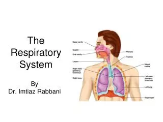
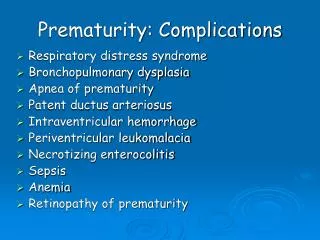
Problem of prematurity (1) • Respiratory • Respiratory distress syndrome (RDS) • Apnea • Bronchopulmonary dysplasia (BPD) • Neurologic • Intraventricular hemorrhage (IVH) • Periventricular leukomalacia (PVL) • Cardiovascular • Hypotension • Patent ductus arteriosus (PDA)
Problem of prematurity (2) • Hematologic • Anemia • Hyperbilirubinemia • Nutritional • Feeding problems • Type, amount, and route of feeding • Gastrointestinal • Necrotizing enterocolitis (NEC) • Metabolic • Acidosis • Hyper- or hypoglycemia • hypocalcemia
Problem of prematurity (3) • Renal • Low GFR • Inability to handle water, solute, and acid loads • Temperature regulation • Hypothermia and hyperthermia • Immunologic • Greater risk for infection • Ophthalmologic • Retinopathy of prematurity (ROP)
Intraventricular hemorrhage (IVH) • In premature infant :--occurs in the gelatinous subependymal germinal matrix --highly vascular area with immature blood vessels • In term infant : --germinal matrix become attenuated and tissue’s vascular support has strengthened.
Intraventricular hemorrhage (IVH) • The incidence of IVH :---60~70% of 500-750 g infants---10~20% of 1000-1500 g infants • 80~90% of cases occur between birth and the 3rd day of life ; 50% occur on the 1st day. • 20~40% of cases progress during the 1st week of life ; delayed hemorrhage may occur in 10~15% of patients after the 1st week of life. • New-onset IVH is rare after the 1st month of life regardless of birth weight.
Predisposing factors for IVH : • --prematurity--RDS--Hypoxic-ischemic or hypotensive injury--reperfusion of damaged vessels--increased or decreased cerebral blood flow--reduced vascular integrity--increased venous pressure--pneumothorax--hypervolemia--hypertension
Clinical manifestations • Diminished or absent Mono reflex • Poor muscle tone • Lethargy • Apnea • Somnolence • Periods of apnea, pallor, or cyanosis • Failure to suck well • Abnormal eye signs • Decreased muscle tone or paralysis • Metabolic acidosis • Shock • Decreased hematocrit or its failure to increase after transfusion
Periventricular leukomalacia (PVL) • A common associated cystic finding • May be due to prenatal or neonatal ischemic or reperfusion injury • The result of necrosis of the periventrucular white matter • Damage to the corticospinal fibers in the internal capsule.
Periventricular leukomalacia (PVL) • Usually asymptomatic until the neurological sequelae of white matter necrosis become apparent in later infancy as spastic diplegia. • May be present at birth but usually occurs later as an early echodense phase (3-10 days of life) followed by the typical echolucent (cystic) phase (14-20 days of life).
Intraventricular hemorrhage (IVH) • Grade I - Germinal matrix hemorrhage (subependymal region or less than 10% of the ventricle; ~35% of IVH) • Grade II - IVH with 10-50% filling of the ventricle (~40% of IVH) • Grade III– more than 50% involvement with dilated ventricles • Grade IV - IVH with extension into the parenchyma
Patent ductus arteriosus (PDA) • Connect the main pulmonary trunk (or proximal left pulmonary artery) with the descending aorta, 5-10 mm distal to the origin of the left subclavian artery • Arising from the distal dorsal sixth aortic arch • Is well developed by the sixth week of gestational age • Is more prevalent in female than male • Is a frequent complication of HMD in preterm infant, in infant born at high altitudes
Normal postnatal closure • First stage : contraction and cellular migration of medial smooth muscle -->result functional closure commonly occurred within 12 hours in full term baby • Second stage : connective tissue formation and replacement of muscle fibers with fibrosis--> ligmentum arteriosum • Both PGE2 and PGI2 relax the ductus arteriosus
Incidence • Prematurity: inverse with GA, PDA is found in about 45 % of infant under 1750g and 80% in infants weighting <1000g • Risk factor: 1.RDS and surfactant treatment 2.Fluid overload 3.Asphyxia 4.Congenital syndrome,congenital heart disease 5.High altitude
Pathophysiology • Ductal constriction is caused by multiple factors :1. oxygen 2. the level of prostaglandin 3. available ductus muscle mass • Within the first hours after birth -> fall in pulmonary vascular resistance and a rise in systemic resistance if PDA opened left to right shunt(+) --> result in increased pulmonary blood flow ,left ventricular volume overload, increased left ventricular end-diastolic volume and pressure ->CHF
Pathophysiology • Renal, mesenteric and cerebral blood flow decreased due to ductal steal • These with moderate and large ducts are prone to the development of pulmonary vascular obstructive disease by 1 year of age or beyond • Preterm infant may develop CHF earlier because of incomplete development of the medial musculature in the small pulmonary arterioles • Among those with RDS, they may be a initial period of improvement as the pulmonary status improves
Clinical findings (Term infants ) • Pulmonary vascular resistance determines the clinical manifestations: • A continuous murmur is heard infrequently • Large PDA has • 1. bounding peripheral pulse pressure, • 2. wide pulse pressure(difference between systolic and diastolic pressure) • 3. hyperactive precordium: due to elevated stroke volume
Clinical findings (Term infants ) • 4. Hypotension particular in these of ELBW • 5. Heart failure in large PDA doesn’t develop until 3 to 6 weeks of age • Associated with pulmonary disease ,left heart obstructive lesion and coarctation of aorta , pulmonary resistance may be high --> right to left shunt --> no murmur
Clinical findings (preterm infants) • 1.The same clinical sign as term baby • 2.However, many preterm baby with large PDA have no murmur • 3.Most will have an increased pressure
Diagnosis • Chest x ray : cardiac enlargement ,pulmonary plethora, a prominent main pulmonary artery and left atrial enlargement • EKG : left ventricular hypertrophy, left atrial hypertrophy • Echocardiography: 1. M-mode : normal LA : Aa ratio in infants is between 0.8-1.0, A ratio > 1.2 suggests left atrial enlargement (in the absence of left ventricular failure or volume overload) 2. 2-D:PDA
Treatment • Term infants : No evidence of cardiovascular embarrassment should be followed and catheter closure or thoracoscopic or surgical diversion • Digoxin and diuretics for PDA with CHF
Preterm infants • 1. Ventilator support and fluid restriction • 2. Indomethacin treatment produces closure in 85% of patients • 3. Prophylactic administration of indomethacin early after birth in very premature infants (<1250 g) decreased the incidence of PDA, CHF, IVH and possibly mortality ----but not routine due to the risk of leukomalacia, decreased renal function, platelet function and NEC
Preterm infants • 4.Ibuprofen(10 mg/kg) may have fewer side effect. Archives of Disease in Childhood: Fetal & Neonatal Edition. 76(3):F179-84, 1997 May. • (ibuprofen did not significantly reduce mesenteric and renal blood flow velocity.) Journal of Pediatrics. 135(6):733-8, 1999 Dec. • 5.Blood transfusion in anemic preterm baby diminishes the left ventricle volume overload and hasten ductus closure by increasing arterial oxygen content
Preterm infants • Early indomethacin treatment (in premature infants with respiratory distress syndrome) improves PDA closure but is associated with increased renal side effects and more severe complications and has no respiratory advantage over late indomethacin administration in ventilated, surfactant-treated, preterm infants <32 weeks' gestational age.(Journal of Pediatrics. 138(2):205-11, 2001 Feb.)
PDA • Coil occlusion is a safe and effective method of percutaneous closure of small to moderate-size (minimum diameter < or = 4 mm) PDAs. • The largest PDA that can be closed with this technique remains to be determined. Journal of Pediatrics. 130(3):447-54, 1997 Mar.
Contraindications for indomethacin • 1.serum creatine >1.7 mg/dl • 2.Frank renal or gastrointestinal bleeding or generalized coagulopathy • 3.NEC • 4.sepsis
Necrotizing enterocolitis • 1.Definition2.Incidence3.Pathology & Pathogenesis4.Clinical manifestations5.Diagnosis6.Management7.Complication
Definition • The most common life-threatening emergency of the gastrointestinal tract in the newbornstage. • An acquired neonatal disorder characterized by various degrees of mucosal or transmural necrosis of the intestine.
Incidence • Decreased birth weight & gestational age incidence & fatility • Rare in term infants. • Overall mortality 20 — 40%. • Neonatal ICU 1— 5 % • No association with or race. • Occures sporadically or in epidemic clusters. • Most involved the distal part of the ileum and the proximal segment of colon.
Pathology & Pathogenesis (1) • Cause : remains unclear but is multifactorial. • No proven cause has been estabilished. • The greatest riskPremature • Interactions between mucosal injury (ischemia, infection, inflammation) and the host’s response to the injury (circulatory, immunologic, inflammatory)
Pathology & Pathogenesis (2) • Clustering of the cases infectious agent (E. Coli., Klebisella, Enterobacter, Salmonella, Coronavirus, Rotavirus, Enterovirus) • No pathogen is identified. • Rarely occures before enteral feeding. • Much less common in infants fed human milk. • Triadintestinal ischemia, oral feeding, pathogenic organisms
Initial ischemic or toxic mucosal damage Loss of mucosal integrity Enteral feedings + Bacterial proliferation Necrosis of the intestine Gas accumulation in the submucosa of bowel wall (penumatosis intestinalis) Transmural necrosis or gangrane Perforation, Sepsis, Death
Clinical manifestations • A variety of signs and symptoms and may be onset insidiously or suddenly. • Usually occurs in the first 2 weeks. • Age of onset is inversely relatede to the gestational age (VLBW 3 month). • First signs : abdominal distension with gastric retention. • 25 % bloody stool • Progress maybe be rapid, but unusually to progress from mild to severe after 72 hr.
Signs and symptoms associated with necrotizing enterocolitis • Gastrointestinal • Abdominal distention • Abdominal tenderness • Feeding intolerance • Delayed gastric emptying • Vomitting • Occult/gross blood stool • Change in stool pattern/ diarrhea • Abdominal mass • Erythema of abdominal wall • Systemic • Lethargy • Apnea/ respiratory distress • Temperature instability • Acidosis • Glucose instability • Poor perfusion/ shock • DIC • Positive results of blood culture
Diagnosis • A very high index of suspicion in treating infants at risk is essential. • Clinical triad: Feeding intolerance, abdominal distention, grossly bloody stools. • Lab studies: CBC, electrolytes, blood culture, stool screening, stool culture,… • Radiologic studies: 1.X-ray of abdomen: Pneumomatosis intestinalis (50-75%) Portal venous gas 2.Hepatic ultrasonography
KUB demonstrating abdominal distention, hepatic portal venous gas(arrow),and bubbly appearance of pneumatosis intestinalis (arrowhead). The latter two signs are pathognomonic for NEC.
Intestinal perforation. Cross-table abdominal roentgenogram in a patient with NEC demonstrating marked distention and massive pneumoperitoneum as evident by the free air below the anterior abdominal wall.
Management • Basic NEC protocol: 1.Nothing by mouth (NPO) 2.Use of a nasogastric tube 3.Antibiotics 4.Monitoring of vital signs & abdominal circumference 5.Removal of the umbilical catheter 6.Monitoring of fluid intake and output 7.Monitoring for gastrointestinal bleeding 8.Laboratory monitoring 9.Septic workup 10.Radiologicstudies
Management byStages • Classified by clinical syndrome (1986 Walsh and Kliegman) • Stage I : Suspected NEC Systemic : Nonspecific, apnea, bradycardia, and temperature instability Gastrointestinal : Increased gastric residuals Occult blood stool Radiographic : Normal or nonspecific Treatment : NPO with antibiotics for 3 days
Stage II A – Mild NEC • Systemic : Nonspecific, similar to stage 1 • Gastrointestinal : Absent bowel sounds and Gross blood stools. • Radiographic : Ileus with dilated loops, focal areas of pneumatosis intestinalis • Treatment : NPO with antibiotics for 10-14 days
Stage II B – Moderate NEC • Systemic: Mild metabolic acidosis and mild thrombocytopenia • Gastrointestinal: Tenderness, abdomianl wall edema,palpable mass • Radiographic: Extensive pneumatosis, portal venous gas, early ascites • Treatment: Similar to stage II B
Stage III A – Advanced NEC • Systemic: Hypotension, bradycardia, respiratory failure, coagulopathy severe metabolic acidosis • Gastrointestinal: Spreading edema, erythema induration of the abdomen • Radiographic: Prominent ascites • Treatment: paracentesis, fluid resuscitation ,inotropic agent support, ventilator support,.
Stage III B – Advanced NEC • Systemic : Deteriorating vital signs, shock, electrolyte imbalance • Gastrointestinal : Perforation of the bowel • Radiographic : Perforation of the bowel • Treatment : Surgical management
Surgical management • Indication for operation: 1.Evidence of intestinal perforation 2.A spersistent, fixed senile loop 3.Erythema of the abdominal wall 4.A palpable mass 5.Brown paracentesis fluid with organisms on Gram stain 6.Failure to response to medical treatment.