Download
1 / 30
340 likes | 700 Vues
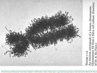
Nucleosomes. Chapter 29. 29.2 The Nucleosome Is the Subunit of All Chromatin. Micrococcal nuclease releases individual nucleosomes from chromatin as 11S particles. Figure 29.2. A nucleosome contains: ∼200 bp of DNA two copies of each core histone (H2A, H2B, H3, and H4). Figure 29.3.

E N D
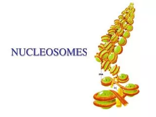
Nucleosomes Chapter 29
29.2 The Nucleosome Is the Subunit of All Chromatin • Micrococcal nuclease releases individual nucleosomes from chromatin as 11S particles. Figure 29.2
A nucleosome contains: • ∼200 bp of DNA • two copies of each core histone (H2A, H2B, H3, and H4) Figure 29.3
DNA is wrapped around the outside surface of the protein octamer. Figure 29.5
29.3 DNA Is Coiled in Arrays of Nucleosomes • >95% of the DNA is recovered in nucleosomes or multimers when micrococcal nuclease cleaves DNA of chromatin. • The length of DNA per nucleosome varies for individual tissues in a range from 154 to 260 bp. Figure 29.7
29.4 Nucleosomes Have a Common Structure • Nucleosomal DNA is divided into the core DNA and linker DNA. • It depends on the DNA’s susceptibility to micrococcal nuclease. • The core DNA is the length of 146 bp that is found on the core particles produced by prolonged digestion with micrococcal nuclease. Figure 29.9
Linker DNA is the region of 8 to 114 bp that is susceptible to early cleavage by the enzyme. • Changes in the length of linker DNA account for the variation in total length of nucleosomal DNA. • H1 is associated with linker DNA and may lie at the point where DNA enters and leaves the nucleosome.
29.5 DNA Structure Varies on the Nucleosomal Surface • DNA is wrapped 1.65 times around the histone octamer. • The structure of the DNA is altered so that it has: • an increased number of base pairs/turn in the middle • a decreased number at the ends
29.6 The Periodicity of DNA Changes on the Nucleosome • ∼0.6 negative turns of DNA are absorbed by the change in bp/turn: • from 10.5 in solution • to an average of 10.2 on the nucleosomal surface • This explains the linking-number paradox.
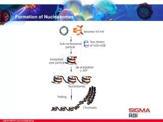
29.7 Organization of the Histone Octamer • The histone octamer has a kernel of an H32-H42 tetramer associated with two H2A-H2B dimers. Figure 29.17
Each histone is extensively interdigitated with its partner. • All core histones have the structural motif of the histone fold. • N-terminal tails extend out of the nucleosome. Figure 29.20
29.8 The Path of Nucleosomes in the Chromatin Fiber • 10 nm chromatin fibers: • are unfolded from 30 nm fibers • consist of a string of nucleosomes Figure 29.23
30 nm fibers have six nucleosomes/turn,. • They are organized into a solenoid. • Histone H1 is required for formation of the 30 nm fiber. Figure 29.25
29.9 Reproduction of Chromatin Requires Assembly of Nucleosomes • Histone octamers are not conserved during replication. • H2A-H2B dimers and H32-H42 tetramers are conserved. Figure 29.28
There are different pathways for the assembly of nucleosomes: • during replication • independently of replication • Accessory proteins are required to assist the assembly of nucleosomes.
CAF-1 is an assembly protein that is: • linked to the PCNA subunit of the replisome • required for deposition of H32-H42 tetramers following replication • A different assembly protein and a variant of histone H3 may be used for replication-independent assembly. Figure 29.30
29.10 Do Nucleosomes Lie at Specific Positions? • Nucleosomes may form at specific positions as the result either of: • the local structure of DNA • proteins that interact with specific sequences • The most common cause of nucleosome positioning is when proteins binding to DNA establish a boundary. Figure 29.31
Positioning may affect: • which regions of DNA are in the linker • which face of DNA is exposed on the nucleosome surface Figure 29.34
29.11 Are Transcribed Genes Organized in Nucleosomes? • Nucleosomes are found at the same frequency when one digests with micrococcal nuclease: • transcribed genes • nontranscribed genes • Some heavily transcribed genes appear to be exceptional cases that are devoid of nucleosomes. Figure 29.36
29.12 Histone Octamers Are Displaced by Transcription • RNA polymerase displaces histone octamers during transcription in a model system. • Octamers reassociate with DNA as soon as the polymerase has passed. • Nucleosomes are reorganized when transcription passes through a gene. Figure 29.38
29.13 Nucleosome Displacement and Reassembly Require Special Factors • Ancillary factors are required both: • for RNA polymerase to displace octamers during transcription • for the histones to reassemble into nucleosomes after transcription Figure 29.41
29.14 Insulators Block the Actions of Enhancers and Heterochromatin • Insulators are able to block passage of any activating or inactivating effects from: • Enhancers • Silencers • LCRs Figure 29.42
Insulators may provide barriers against the spread of heterochromatin. Figure 29.43
29.15 Insulators Can Define a Domain • Insulators are specialized chromatin structures that have hypersensitive sites. • Two insulators can protect the region between them from all external effects. Figure 29.44
29.16 Insulators May Act in One Direction • Some insulators have directionality; • They may stop passage of effects in one direction but not the other. Figure 29.46
29.17 Insulators Can Vary in Strength • Insulators can differ in how effectively they block passage of an activating signal.
29.18 DNAase Hypersensitive Sites Reflect Changes in Chromatin Structure • Hypersensitive sites are found at the promoters of expressed genes. • They are generated by the binding of transcription factors that displace histone octamers. Figure 29.51
29.19 Domains Define Regions That Contain Active Genes • A domain containing a transcribed gene is defined by increased sensitivity to degradation by DNAase I. Figure 29.52
29.20 An LCR May Control a Domain • An LCR: • is located at the 5′ end of the domain • consists of several hypersensitive sites Figure 29.54
29.21 What Constitutes a Regulatory Domain? • A domain may have: • an insulator • an LCR • a matrix attachment site • transcription unit(s) Figure 29.54