Download
1 / 1
10 likes | 419 Vues
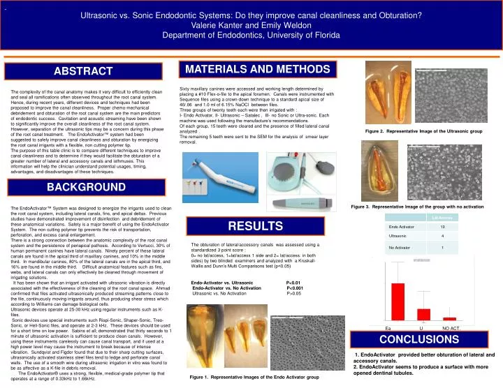
Ultrasonic vs. Sonic Endodontic Systems: Do they improve canal cleanliness and Obturation? Valerie Kanter and Emily Weldon Department of Endodontics, University of Florida. . MATERIALS AND METHODS. ABSTRACT.

E N D
Ultrasonic vs. Sonic Endodontic Systems: Do they improve canal cleanliness and Obturation? Valerie Kanter and Emily Weldon Department of Endodontics, University of Florida . MATERIALS AND METHODS ABSTRACT Sixty maxillary canines were accessed and working length determined by placing a #10 Flex-o-file to the apical foramen. Canals were instrumented with Sequence files using a crown-down technique to a standard apical size of 40/.06 and 1.0 ml of 6.15% NaOCl between files. Three groups of twenty teeth each were then irrigated with : I- Endo Activator, II- Ultrasonic – Satalec , III- no Sonic or Ultra-sonic. Each machine was used following the manufacture’s recommendations. Of each group, 15 teeth were cleared and the presence of filled lateral canal analyzed . The remaining 5 teeth were sent to the SEM for the analysis of smear layer removal. The complexity of the canal anatomy makes it very difficult to efficiently clean and seal all ramifications often observed throughout the root canal system. Hence, during recent years, different devices and techniques had been proposed to improve the canal cleanliness. Proper chemo-mechanical debridement and obturation of the root canal system are the main predictors of endodontic success. Cavitation and acoustic streaming have been shown to significantly improve the overall cleanliness of the root canal system. However, separation of the ultrasonic tips may be a concern during this phase of the root canal treatment. The EndoActivator™ system had been suggested to safely improve canal cleanliness and obturation by energizing the root canal irrigants with a flexible, non cutting polymer tip. The purpose of this table clinic is to compare different techniques to improve canal cleanliness and to determine if they would facilitate the obturation of a greater number of lateral and accessory canals and isthmuses. This information will help the clinician understand potential usages, timing, advantages, and disadvantages of these techniques. Figure 2. Representative Image of the Ultrasonic group BACKGROUND The EndoActivator™ System was designed to energize the irrigants used to clean the root canal system, including lateral canals, fins, and apical deltas. Previous studies have demonstrated improvement of disinfection and debridement of these anatomical variations. Safety is a major benefit of using the EndoActivator System. The non cutting polymer tip prevents the risk of transportation, perforation, and excess canal enlargement. There is a strong connection between the anatomic complexity of the root canal system and the persistence of periapical pathosis. According to Vertucci, 30% of human permanent canines have lateral canals. Ninety percent of these lateral canals are found in the apical third of maxillary canines, and 10% in the middle third. In mandibular canines, 80% of the lateral canals are in the apical third, and 16% are found in the middle third. Difficult anatomical features such as fins, webs, and lateral canals can only effectively be cleaned through movement of irrigating solutions. It has been shown that an irrigant activated with ultrasonic vibration is directly associated with the effectiveness of the cleaning of the root canal space. Ahmad confirmed that files activated ultrasonically produced streaming patterns close to the file, continuously moving irrigants around, thus producing shear stress which according to Williams can damage biological cells. Ultrasonic devices operate at 25-30 kHz using regular instruments such as K-files. Sonic devices use special instruments such Rispi-Sonic, Shaper-Sonic, Treo-Sonic, or Heli-Sonic files, and operate at 2-3 kHz. These devices should be used for a short time on low power. Sabins et all; demonstrated that thirty seconds to 1 minute of ultrasonic activation is sufficient to produce clean canals. However, using these instruments carelessly can cause canal transport, and if used at a high power level may cause the instrument to break because of intense vibration. Sundqvist and Figdor found that due to their sharp cutting surfaces, ultrasonically activated stainless steel files tend to ledge and perforate canal walls. The use of a smooth wire during ultrasonic irrigation in vitro was found to be as affective as a K-file in debris removal. The EndoActivator® uses a strong, flexible, medical-grade polymer tip that operates at a range of 0.33kHz to 1.66kHz. Figure 3. Representative Image of the group with no activation RESULTS The obturation of lateral/accessory canals was assessed using a standardized 3 point score : 0= no lat/access, 1=lat/access 1 side and 2= lat/access. in both sides) by two blinded examiners and analyzed with a Kruskall-Wallis and Dunn’s Multi Comparisons test (p<0.05) Endo-Activator vs. Ultrasonic P<0.01 Endo-Activator vs. No Activation P<0.001 Ultrasonic vs. No Activation P>0.05 Ea U NO ACT. CONCLUSIONS 1. EndoActivator provided better obturation of lateral and accessory canals. 2. EndoActivator seems to produce a surface with more opened dentinal tubules. Figure 1. Representative Images of the Endo Activator group