Download
1 / 55
550 likes | 644 Vues
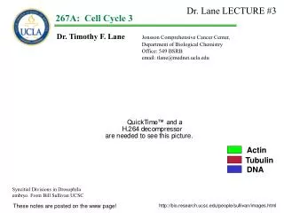
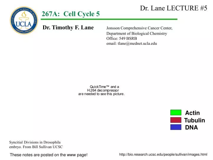
Dr. Lane LECTURE #5. 1. 267A: Cell Cycle 5. Dr. Timothy F. Lane Jonsson Comprehensive Cancer Center, Department of Biological Chemistry Office: 549 BSRB email: tlane@mednet.ucla.edu. Actin. Tubulin. DNA. Syncitial Divisions in Drosophila embryo. From Bill Sullivan UCSC.

E N D
Dr. Lane LECTURE #5 1 267A:Cell Cycle 5 Dr. Timothy F. Lane Jonsson Comprehensive Cancer Center, Department of Biological Chemistry Office: 549 BSRB email: tlane@mednet.ucla.edu Actin Tubulin DNA Syncitial Divisions in Drosophila embryo. From Bill Sullivan UCSC These notes are posted on the www page! http://bio.research.ucsc.edu/people/sullivan/images.html
Last time: We talked in even greater length about the regulation of the cell cycle by cyclins and cdks: i. We focused on how regulatory signals are terminated so that the cell can enter subsequent stages, or a new cell cycle. ii. Focusing on M, we identified a cycB degrading activity called APC. iii. We found that securins were also degraded by APC, but much earlier. iv. that turnover could be very precisely programmed by different E3 ligases (CDH1 for cycB and cdc20 for securins). v. We identified sequences within the targets (cycB and cdc20) that provided specific interaction surfaces for binding of APCCDH1 or APCCDC20) vi. We found that APCCDH1 function continued into G1, and that G1 cyclins were required to terminate APCCDH1 function. We have now discussed 4 levels of regulation for cyclins and cdks, including: i. Transcriptional control of synthesis (pRB represses cycE) ii. Phosphorylation (wee1 etc) / dephosphorylation (cdc25) iii. binding to CKIs (Far1, Sic, p15) iv. turnover by APC.
Goals: The context will be Mitosis. How M-phase is regulated/ended! Examine how cyclins are turned over in the context of M
Mitosis: WHAT DIRECTS APC ACTIVITY: The APCcdc20 complex peaks in activity in M, and is responsible for Pds1p/securin proteolysis and anaphase onset. APCCdh1 cyclosome activity peaks in Anaphase/G1, and is responsible for promoting Clb2p proteolysis and consequent G1 entry from M Pds1/Cut2/securin Nasmyth, 1999 TIBS 24:98
CDH1 separase LAST TIME: Cell cycle dependent protein degradation during Mitosis APCcdc20 complex responsible for Pds1p/securin proteolysis and anaphase onset. APCCdh1 complex responsible for cycB proteolysis and G1 entry. G1 S APC Cyclin B G2 M CDH1 o o o o o o Cdc 20 securin APC
THE G1 CYCLINS ARE PREPARED FOR PROTEASOME DEGRADATION BY A DISTINCT UBIQUITINATION MECHANISM –THE SCF COMPLEX Mitogen signal for G0 cells Cyclin D1 cdk4 R.P. Cyclin E Go G1 Cyclin B M cdk2 cdk2 Cyclin A G2 cdk2 S
THE G1 CYCLINS ARE PREPARED FOR PROTEASOME DEGRADATION BY A DISTINCT UBIQUITINATION MECHANISM –THE SCF COMPLEX Cdc34 mutants arrest at the G1/S transition, Cdc34p encodes a ubiquitination enzyme1. Cln2 cdc28 Go G1 M G2 S 1Goebl et al 1988 Science 241:1331
THE G1 CYCLINS ARE PREPARED FOR PROTEASOME DEGRADATION BY A DISTINCT UBIQUITINATION MECHANISM –THE SCF COMPLEX Cdc34 mutants arrest at the G1/S transition, Cdc34p encodes a ubiquitination enzyme1. Recombinant cdc34 protein has endogenous ubiquitin-conjugating activity with histone 2A1 Cln2 cdc28 Go G1 M G2 S 1Goebl et al 1988 Science 241:1331
Ub Ub Ub Ub THE G1 CYCLINS ARE PREPARED FOR PROTEASOME DEGRADATION BY A DISTINCT UBIQUITINATION MECHANISM –THE SCF COMPLEX Cln2 (recomb) Cdc34 mutants arrest at the G1/S transition, Cdc34p encodes a ubiquitination enzyme1. p cdc28 p cln2 Recombinant cdc34 protein has endogenous ubiquitin-conjugating activity with histone 2A1 Extract from cyclin depleted, G1 arrested S.c. + Cdc34p ubiquitinates Cln2 and leads to Cln2 destruction2. The Cln2 in the complex is phosphorylated! In these in vitro reactions, an additional modified form of Cln2-P is observed. This Cln2-P modified form can be precipitated with anti-ubiquitin. cdc34 Ub Ub p Ub p cln2 The ubiquitination occurs in extracts of wt. cells, but not extracts of cdc34 t.s. cells. Moreover, ubiquitination did not occur in cdc28 t.s. extracts2. 1Goebl et al 1988 Science 241:1331 2Deshaies et al 1995 EMBO J. 14:303
Cln3 Beta -galactosidase THE G1 CYCLINS ARE PREPARED FOR PROTEASOME DEGRADATION BY A DISTINCT UBIQUITINATION MECHANISM –THE SCF COMPLEX Cln3 is phosphorylated by Cdc28/p34, and is then ubiquitinated by a Cdc34-dependent step, leading to Cln3 degradation. As with Cln2, Cln3 is not degraded in cdc28 t.s. mutants! Expt. Create a Cln3/gal fusion protein Expressed in wt or cdc34 mutant yeast. The proteins were labeled for five minutes with radioactive amino acids, then the cells were chased with cold amino acids. At the times shown, Cln3-gal was immunoprecipitated and subjected to electrophoresis and autoradiography Wild-type yeast strain cdc34 mutant yeast strain “[35S]-Met” amino acid pulse cold amino acid chase i.p. Electrophoresis Yaglom et al, 1995 Mol. Cell. Biol. 15:731
THE G1 CYCLINS ARE PREPARED FOR PROTEASOME DEGRADATION BY A DISTINCT UBIQUITINATION MECHANISM –THE SCF COMPLEX Cln3 is phosphorylated by Cdc28/p34, and is then ubiquitinated by a Cdc34-dependent step, leading to Cln3 degradation. As with Cln2, Cln3 is not degraded in cdc28 t.s. mutants! Expt. Create a Cln3/gal fusion protein Expressed in wt or cdc34 mutant yeast. The proteins were labeled for five minutes with radioactive amino acids, then the cells were chased with cold amino acids. At the times shown, Cln3-gal was immunoprecipitated and subjected to electrophoresis and autoradiography Cdc34 Cln3-beta gal remaining w.t. RESULT: Cln3 is very stable in cells lacking the cdc34 ubiquitin ligase 15 30 45 60 Chase time (min) Yaglom et al, 1995 Mol. Cell. Biol. 15:731
p p CKIs are PREPARED FOR PROTEASOME DEGRADATION BY A CDC34 DEPENDENT PROCESS SIC1 degradation in G1 is necessary for entry into S phase Recall from Lecture #3: To enter S, p40/Sic1 protein is phosphorylated and then degraded, releasing the inhibition of the CDK activity. cdc28 cdc28 SIC 1 SIC 1 p p Clb2 Clb2 G1 S Histone Histone Histone Histone 1Mendenhall, 1993 Science 259:216 2Schwob et al, 1994, Cell 79: 233
cdc34 P P P P P P P CKIs are PREPARED FOR PROTEASOME DEGRADATION BY A CDC34 DEPENDENT PROCESS • The transition from G1 to S should involve a rapid initiation of DNA synthesis • But must occur only after a series of events in G1 has taken place. • Can not be dependent on small errors in the level of phosphorylation of Sic1. • In fact, six out of nine phosphorylation sites on Sic1 must be phosphorylated before • ubiquitination/degradation becomes effective. • This results in a delayed, but sharp initiation of Sic1 degradation, • activation of cdc2/S cyclin protein kinase activity, and the G1/S transition. cdc28 Cln2 SIC 1 SIC 1 DEGRADATION G1 S Nash et al, 2001 Nature 414:514-521
cdc34 cdc4 ub CDC34 DEPENDENT UBIQUITINATION REQUIRES THE SCF complex Several other gene products are required for the G1/S transition When mutated – some produce phenotypes like cdc34 (i.e., arrest at G1/S). These include: skp1, cdc53 and cdc4. The SCF complex is required to recognize pSic1 protein and catalyze its ubiquitination. Skowyra et al expressed recombinant forms of all these proteins in an insect cell system and used co-immunoprecipitation procedures to demonstrate that four proteins – Cdc34, Cdc4, Cdc53, and Skp1 – could form a macromolecular complex1. Feldman et al demonstrated that this complex, in the presence of ubiquitin and the E1 and E2 enzymes, can ubiquitinate phosphorylated Sic12. The cdc4 protein recognizes the multi-phosphorylated Sic1 protein, and causes its binding to the ubiquitinating complex Sic 1 p p Skp1 Cdc53 1Skowyra et al 1997 Cell 91:209 2Feldman et al 1997, Cell 91: 221
cdc34 cdc4 ub ub ub ub ub ub G1 Cyclin S-phase Cyclin S-phase Cyclin cdc28 cdc28 cdc28 Sic 1 Sic 1 p p Inactive kinase Skp1 Cdc53 S-phase Cyclin proteasome S-phase Cyclin cdc28 cdc28 Sic 1 p p active kinase
p p p p p p p p cyclin CDC34 DEPENDENT UBIQUITINATION targets multiple targets p cdk cdk WEE1 cyclin Phosp. Thr 161 cyclin WEE1 Dephosp. Thr 161 p cdc25 p cdk cdc25 cyclin ACTIVE CDK Degrade cyclin CKI CKI cdk cdk p cyclin
cdc34 cdc4 ub Structure of SCF cdc4 associates with Skp1 through a motif on Cdc4 that is called an F-box. The name SCF complex (for Skp1, Cdc53, F-box protein). F-box cdc4 Sic 1 p p Skp1 Cdc53 Bai et al 1996, Cell 86:263 Skowra et al 1997, Cell 91:209
Structure of SCF cdc4 associates with Skp1 through a motif on Cdc4 that is called an F-box. The name SCF complex (for Skp1, Cdc53, F-box protein). Bai et al identified a number of proteins that contain F-box domains. They suggested that different F box domain proteins might select alternative targets for SCF. Skowra et al then demonstrated that this is the case: F-box cdc4 F box-2 F box-3 Bai et al 1996, Cell 86:263 Skowra et al 1997, Cell 91:209
cdc34 ub ub ub ub ub ub Structure of SCF cdc4 associates with Skp1 through a motif on Cdc4 that is called an F-box. The name SCF complex (for Skp1, Cdc53, F-box protein). Cln2 Cln2 p p Bai et al identified a number of proteins that contain F-box domains. They suggested that different F box domain proteins might select alternative targets for SCF. GRR1 Skp1 Cdc53 Skowra et al then demonstrated that this is the case: F-box protein, Grr1, associates with SCF components Skp1, Cdc53, and Cdc34. SCFGrr1not bind to p-Sic1, but does bind to p-Cln2 & p-Cln3 It appears that the SCF complex – interacts with a variety of F-box proteins, via the Skp1 recognition site. The F-box containing subunits target the SCF ubiquitination activity to different substrates. Cln2 p p Bai et al 1996, Cell 86:263 Skowra et al 1997, Cell 91:209
Structure of SCF: Selecting multiple targets: SCFGRR1 targets G1 cyclins for proteosomal turnover: SCFCDC4 targets Sic1 for proteosomal turnover:
p p Structure of SCF: Selecting multiple targets: Wee1 is also degraded by an SCF Cdc34 mechanism. Recall that wee1 (a kinase that phosphorylates p34cdc2) in S. pombe must be inactivated for cells to move from G2 into M, wee1 protein is phosphorylated and degraded. wee1 ATP Cdc2 p34 Cdc2 p34 cdc 13 cdc 13 ATP HISTONE H1 H1-P Michael and Newport 1998, Science 282: 1886
p p Structure of SCF: Selecting multiple targets: Wee1 is also degraded by an SCF Cdc34 mechanism. Recall that wee1 (a kinase that phosphorylates p34cdc2) in S. pombe must be inactivated for cells to move from G2 into M, wee1 protein is phosphorylated and degraded. Expt: Using an oocyte extract system, it was demonstrated that a dominant-negative cdc34 mutant can block wee1 degradation. Thus, in addition to ubiquitinating G2 phase cyclins and CKIs, an SCF complex regulates entry into mitosis by initiating wee1 protein degradation. What F-box protein targets wee1 kinase for ubiquitination? wee1 wee1 DEGRADATION cdc34dn Michael and Newport 1998, Science 282: 1886
cdc34 ub What F-box protein targets Wee1 for destruction? Ayad et al used a biochemical search to find new substrates for APCCDH1. They in vitro translated pools of cDNAs, mixed them with: [mitotic extracts + CDH1] vs [mitotic extracts without CDH1], looked for proteins degraded in the presence of CDH1. RESULT: One of the substrates was called Tome-1. Wee1 p p ??? Skp1 Cdc53 Ayad et al 2003, Cell 113:101-113
G2/M transition What F-box protein targets Wee1 for destruction? As expected for a APCCDH1 substrate, Tome-1 is degraded in a cell cycle dependent manner similar to cyclin B (A) The Tome-1 sequence contains what looks like an F-box. Tome-1 co-IPs with Skp-1 and Cul-1. If Tome-1 is reduced by using a siRNA - wee1 degradation is inhibited. The experiment was done by pulse labeling cells with [35S]cys after incubation with siRNA, then doing IP’s at various times after labeling. Non-mitotic extracts can be stimulated to enter M, and activate CDK activity on histones. If Tome-1 is depleted from extracts with anti-Tome-1, and the extracts are then stimulated. Tome-1 depleted extracts enter mitosis later. G1/S synchronized cells. 100 Tome-1 siRNA Percent wee1 50 Control RNA 50 150 Time (min) 60 40 H1 kinase activity Tome-1 depleted extract 20 Time (min) 50 100 Ayad et al 2003, Cell 113:101-113
cdc34 ub ub ub ub ub ub What F-box protein targets Wee1 for destruction? wee1 Tome-1 is named for trigger of mitotic entry. Tome-1 helps to regulate the transition from G2 into M by acting as an F-box protein leading to the degradation of wee1, shifting the wee1/cdc25 balance in the direction of cdc25. This causes dephosphorylation of cdc2 and activation of the cdc2/cyclin B kinase activity required for mitosis. However, Tome-1 is degraded by an APC-dependent process, so that wee1 can be built back up in the next cycle, preventing premature mitosis. TF: Tome-1 connects the APC and SCF protein degradation cycles. From the yeast data base, there are over 20 “F-box” containing proteins that are candidates for SCF-regulatory molecules that control cell cycle transitions of various types, by promoting ubiquitination and degradation Koepp et al 1999 Cell 97:431 wee1 p p wee1 p p Tome-1 Skp1 Cdc53 wee1 p p
p p p p p p p p cyclin cyclin p cdk cdk WEE1 cyclin Phosp. Thr 161 cyclin WEE1 Dephosp. Thr 161 p cdc25 p cdk cdc25 ACTIVE CDK Degrade cyclin CKI CKI CKI cdk cdk p cyclin
SCF SCF SCF CDH1 Grr1 Cln2 separase cdc4 Sic1 CDH1 APC G1 S Tome-1 APC Cyclin B G2 M wee1 CDH1 o o o Tome-1 o o o Cdc 20 securin APC
The SCF complex The APC/cyclosome
1) Replication of the DNA must occur once, for each gene in a cell cycle, and G2 phase cells are fused to S phase cells – the G2 phase cell DNA does not replicate, despite the presence of a factor that can cause G1 phase cell DNA to replicate. Is there a factor that “licenses ” DNA replication for G1 phase cells? a factor that is removed after replication in S phase? If so, the DNA must be re-licensed before the next S phase. DNA Replication Licensing
DNA Replication Licensing DNA replication occurs at origins of replication (ORC). The origin replication complex, or ORC, binds to replication origins. The ORC is “always” associated with origins of replication.
DNA Replication Licensing “Licensing” occurs as the cells complete mitosis: Cdc18p, Cdt1p, and the MCM proteins (the mini-chromosome maintenance oligomer) are loaded onto the ORC, licensing it for replication.
DNA Replication Licensing Then the MCM is added Unlicensed origin
DNA Replication Licensing The licensed replication complex is activated for DNA replication at the G1/S transition. In yeast, this activation requires Cdc28/clb5 or Cdc28/clb6 –“the S phase cyclins”. In mammalian cells, this activation requires Cdk2-cyclinE. The activation of the licensed replication complex by the CDK results in additional proteins binding to the origin. The ORC is left at the origin, while the assembled, activated replication machinery proceeds down the DNA. The functional target(s) of the CDK required to activate a licensed origin are not yet known. A cdk/S phase cyclin reaction(s) is required to initiate DNA synthesis
DNA Replication Licensing After DNA replication is initiated, Cdc6/18 is then destroyed by an SCFcdc4 ubiquitination and proteasome degradation. Unlicensed origin
HIGH THROUGHPUT SCREENING, GENOMICS, BIO INFORMATICS AND PROTEOMICS COME TO CELL CYCLE ANALYSIS High throughput screens: Do the temperature sensitive screens of Hartwell and Nurse identify all cell cycle genes? Stevenson et al used over-expression of yeast genomic fragments and cDNA libraries, in which the genes are over-expressed from the GAL1 promoter, to look for genes that would cause cell cycle arrest in S. cerevisiae when the cells are shifted to galactose. Using high-throughput screening, 150,000 colonies were screened, 179 genes/ORFs were identified that modulate the cell cycle. Cells were analyzed by flow cytometry after 6-8 hours in the presence of galactose. Strains in the left column are shifted toward a 1C amount of DNA relative to cells containing a control plasmid, cells in the right column are shifted toward a 2C content of DNA. 43% of the known genes cloned had previously been associated with cell cycle. 19 new ORFs were identified that modulated cell cycle. Seven of these ORFs cause accumulation in M phase, when examined microscopically. Stevenson et al 2001,PNAS 98: 3946
Control morphology studies and flow cytometry patterns for S. Cerevisiae Strains in the left column are shifted toward a 1C amount of DNA relative to cells containing a control plasmid, cells in the right column are shifted toward a 2C content of DNA. 43% of the known genes cloned had previously been associated with cell cycle. 19 new ORFs were identified that modulated cell cycle. Seven of these ORFs cause accumulation in M phase, when examined microscopically. Stevenson et al 2001,PNAS 98: 3946
Control flow cytometry patterns for S. Cerevisiae 150 thousand colonies were screened. 179 colonies were isolated in which over expression of gal-driven genomic fragments or cDNAs caused cell cycle arrest. Stevenson et al 2001,PNAS 98: 3946
Strains in the left column are shifted toward a 1C amount of DNA relative to cells containing a control plasmid. Stevenson et al 2001,PNAS 98: 3946
Strains in the right column are shifted toward a 2C amount of DNA relative to cells containing a control plasmid. Stevenson et al 2001,PNAS 98: 3946
HIGH THROUGHPUT SCREENING, GENOMICS, BIO INFORMATICS AND PROTEOMICS COME TO CELL CYCLE ANALYSIS High throughput screens: Spellman et al. synchronized yeast cells three ways (with mating factor, using the cdc15 ts mutant, and using an elutriation rotor). They extracted RNA at various times after release from the cell cycle blocks, and then performed microarray analyses of mRNA levels for the whole yeast genome, versus RNA from exponentially growing cells. They first used “phase analysis,” in which they plotted the peak time of expression for these genes. They found 800 cell cycle regulated genes; 300 in G1, 71 in S, 121 in G2, 195 in M and 113 “M/G1” genes. They compared the upstream promoter sequences of the genes, and found increased presence of appropriate transcription factor response elements. They then used “cluster analysis”, which “sorts through all the data to find the pairs of genes that behave most similarly in each experiment, and then progressively adds other genes to the initial pairs to form clusters of apparently co-regulated genes.” RESULT: Nine clusters were identified (3 in G1, 2 in S, 1 in M, and 3 in M/G1). About half the 800 cell cycle regulated genes are in these nine clusters. Spellman et al. 1998, Mol Biol Cell. 9: 3273
HIGH THROUGHPUT SCREENING, GENOMICS, BIO INFORMATICS AND PROTEOMICS COME TO CELL CYCLE ANALYSIS Analysis of the 5’ regions of the genes in a cluster shows that such genes share common promoter elements. For example, within the G1 group of genes is the “CLB2 cluster” of 76 genes . CRCGAAA ACGCGN 52% of the G1 “CLB2 cluster” genes have this sequence in the promoter; only 13% of all yeast genes have this sequence. 58% of the G1 “CLB2 cluster” genes have this sequence in the promoter; only 6% of all yeast genes have this sequence. Spellman et al. 1998, Mol Biol Cell. 9: 3273
HIGH THROUGHPUT SCREENING, GENOMICS, BIO INFORMATICS AND PROTEOMICS COME TO CELL CYCLE ANALYSIS 76 genes are in the G1 “CLB2” cluster SBF MBF Swi4 Swi6 Mbp1 Swi6 CRCGAAA ACGCGN 52% of the G1 “CLB2 cluster” genes have this sequence in the promoter; only 13% of all yeast genes have this sequence. 58% of the G1 “CLB2 cluster” genes have this sequence in the promoter; only 6% of all yeast genes have this sequence. Spellman et al. 1998, Mol Biol Cell. 9: 3273
HIGH THROUGHPUT SCREENING, GENOMICS, BIO INFORMATICS AND PROTEOMICS COME TO CELL CYCLE ANALYSIS All the data from Spellman et al, 400,000 data points, are freely available on the web for investigation and manipulation by any informatics aficionados. SBF MBF Swi4 Swi6 Mbp1 Swi6 CRCGAAA ACGCGN However, all is not necessarily well….. Shedden and Cooper [Nuc. Acids. Res. 30:2920 (2002)] have reviewed this paper and conclude 1) “…when the degree of cyclicity for genes in different experiments are compared, a large degree of non-reproducibility is found.” 2) “Specific genes can show a wide range of cyclical behavior between different experiments; a gene with high cyclicity in one experiment can show essentially no cyclicity in another experiment.” Spellman et al. 1998, Mol Biol Cell. 9: 3273
HIGH THROUGHPUT SCREENING, GENOMICS, BIO INFORMATICS AND PROTEOMICS COME TO CELL CYCLE ANALYSIS Cell cycle gene profiling has also been extended to mammalian cells. Cho et al synchronized fibroblasts by a double thymidine block and analyzed mRNA profiles every two hours by microarray, for cyclically expressed genes. Twelve “patterns” were identified, which included 731 transcripts. An attempt to classify these patterns into groups of genes involved in related functions is described. Whitfield et al synchronized Hela cells by (1) double thymidine block, (2) high thymidine followed by nocodazole or (3) mitotic collection and pooling on ice. Cells were released, RNA was isolated at various times, and microarray analysis was performed. They identified ~850 cell cycle regulated genes. Extensive temporal correlations and functional classifications are provided. Cho et al 2001, Nature Genetics 27: 48 Whitfield et al 2002, Mol. Biol. Cell 13: 1977
BUT THERE ARE REAL PROBLEMS: Whitfield et al compared the ~700 genes from the Cho et al paper with 595 genes in their own study. Only 96 genes were identified as cell cycle regulated in both studies!! They comment “We have no ready explanation of this difference in results, except to note that there are differences in the cell lineage, the microarray technology, and in the analysis methods…. We suspect that the most significant differences may well be in the degree of synchrony achieved…” 591 GENES 731 GENES CHO cells HeLa cells Only 96 cell cycle regulated genes overlap in these two studies!
BUT THERE ARE REAL PROBLEMS: Whitfield et al compared the ~700 genes from the Cho et al paper with 595 genes in their own study. Only 96 genes were identified as cell cycle regulated in both studies!! They comment “We have no ready explanation of this difference in results, except to note that there are differences in the cell lineage, the microarray technology, and in the analysis methods…. We suspect that the most significant differences may well be in the degree of synchrony achieved…” Again, all is not well….. Shedden and Cooper have also reviewed the data from the Cho et al article. They conclude that the cyclic variations observed do not support the proposal that there are numerous cell-cycle-specifically expressed genes in human cells.” 591 GENES 731 GENES CHO cells HeLa cells Only 96 cell cycle regulated genes overlap in these two studies! Shedden and Cooper 2002, PNAS 99: 4379
HIGH THROUGHPUT SCREENING, GENOMICS, BIO INFORMATICS AND PROTEOMICS COME TO CELL CYCLE ANALYSIS Genomic Analysis of Transcription factor Binding (ChIP on CHIP). A variety of transcription factors have been shown to bind to the upstream regions of members of the various “clusters” of cell-cycle regulated genes in S. cerevisiae. G1 phase of the yeast cell cycle bind either SBF, a heterodimer of Swi4p and Swi6p, or MBF, a heterodimer of Mbp1p and Swi6p. SBF MBF Swi4 Swi6 Mbp1 Swi6 CRCGAAA ACGCGN Iyer et al 2001, Nature 409: 533
ChIP positive genes All intergenic sequences Sequence SBF MBF CRCGAAA 10% 49% --- ACGCGN 20% --- 59% HIGH THROUGHPUT SCREENING, GENOMICS, BIO INFORMATICS AND PROTEOMICS COME TO CELL CYCLE ANALYSIS Genomic Analysis of Transcription factor Binding (ChIP on CHIP). Iyer et al used chromatin immunoprecipitation assays to identify genes that bind SBF and MBF targets. They identified about 200 candidate target genes. 49% of the Swi4 ChIP targets contain CRCGAAA, the SBF target; only 10% of all intergenic regions have this sequence. 59% of the MBF putative targets have the ACGCGN sequence; only 20% of all intergenic regions have this sequence. Of particular interest: “Not every promoter bound by SBF or MBF in vivo contained a recognizable consensus binding site. Moreover, most of the coding sequences and many of the promoters that contain the consensus sequences show no evidence of binding to these factors in vivo.” Using the data of Spellman et al, they found that 66% of the genes identified by ChIP with Swi4p,Swi6 or Mbp1 are cell cycle regulated, as opposed to 13% of all yeast genes. Iyer et al 2001, Nature 409: 533