Download
1 / 1
10 likes | 144 Vues
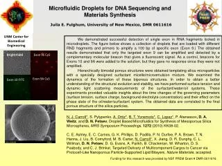
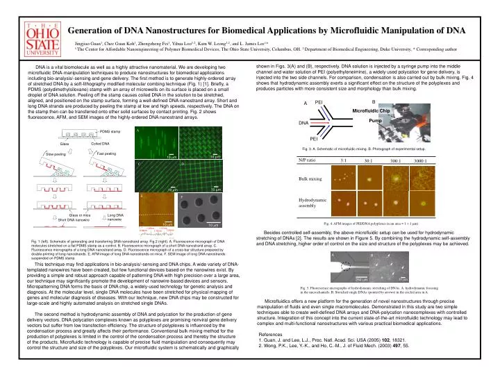
A. N/P ratio. 3:1. 30:1. 300:1. 3000:1. 10 m. Bulk mixing. Hydrodynamic assembly. Fig. 4. AFM images of PEI/DNA polyplexes (scan area = 1 1 m). Generation of DNA Nanostructures for Biomedical Applications by Microfluidic Manipulation of DNA.

E N D
A N/P ratio 3:1 30:1 300:1 3000:1 10 m Bulk mixing Hydrodynamic assembly Fig. 4. AFM images of PEI/DNA polyplexes (scan area = 1 1 m) Generation of DNA Nanostructures for Biomedical Applications by Microfluidic Manipulation of DNA Jingjiao Guan1, Chee Guan Koh1, Zhengzheng Fei1, Yihua Loo1,2, Kam W. Leong1,2, and L. James Lee1* 1.The Center for Affordable Nanoengineering of Polymer Biomedical Devices, The Ohio State University, Columbus, OH. 2.Department of Biomedical Engineering, Duke University, * Corresponding author shown in Figs. 3(A) and (B), respectively. DNA solution is injected by a syringe pump into the middle channel and water solution of PEI (polyethyleneimine), a widely used polycation for gene delivery, is injected into the two side channels. For comparison, condensation is also carried out by bulk mixing. Fig. 4 shows that hydrodynamic assembly exerts a significant effect on the structure of the polyplexes and produces particles with more consistent size and morphology than bulk mixing. • DNA is a vital biomolecule as well as a highly attractive nanomaterial. We are developing two microfluidic DNA-manipulation techniques to produce nanostructures for biomedical applications including bio-analysis/-sensing and gene delivery. The first method is to generate highly-ordered array of stretched DNA by a soft-lithography modified molecular combing technique (Fig. 1) [1]. Briefly, a PDMS (polydimethylsiloxane) stamp with an array of microwells on its surface is placed on a small droplet of DNA solution. Peeling off the stamp causes coiled DNA in the solution to be stretched, aligned, and positioned on the stamp surface, forming a well-defined DNA nanostrand array. Short and long DNA strands are produced by peeling the stamp at lowand high speeds, respectively. The DNA on the stamp then can be transferred onto other solid surfaces by contact printing. Fig. 2 shows fluorescence, AFM, and SEM images of the highly-ordered DNA nanostrand arrays. B PEI A Microfluidic Chip Pump DNA B PDMS stamp PEI Coiled DNA Glass Fig. 3. A. Schematic of microfluidic mixing. B. Photograph of experimental setup. Fast peeling Slow peeling 10 m D C 10 m 10 m E F Long DNA nanowire Glass or mica Short DNA nanowire 10 m 10 m 1.21nm 1.26nm 1.26nm 0.94nm 1.18nm Besides controlled self-assembly, the above microfluidic setup can be used for hydrodynamic stretching of DNAs [2]. The results are shown in Figure 5. By combining the hydrodynamic self-assembly and DNA stretching, higher order of control on the size and structure of the polyplexes may be achieved. Fig. 1 (left). Schematic of generating and transferring DNA nanostrand array. Fig.2 (right). A. Fluorescence micrograph of DNA molecules stretched on a flat PDMS stamp as a control. B. Fluorescence micrograph of a short DNA nanostrand array. C. Fluorescence micrographs of a long DNA nanostrand array. D. Fluorescence micrograph of a cross-bar structure prepared by double-printing of long nanostrands. E. AFM image of long DNA nanostrands on mica. F. SEM image of long DNA nanostrands suspended on PDMS stamp. A B This technique may find applications in bio-analysis/-sensing and DNA chips. A wide variety of DNA-templated nanowires have been created, but few functional devices based on the nanowires exist. By providing a simple and robust approach capable of patterning DNA with high precision over a large area, our technique may significantly promote the development of nanowire-based devices and sensors. Micropatterning DNA forms the basis of DNA chip, a widely-used technology for genetic analysis and diagnosis. At the molecular level, single DNA molecules have been stretched for physical mapping of genes and molecular diagnosis of diseases. With our technique, new DNA chips may be constructed for large-scale and highly automated analysis on stretched single DNAs. The second method is hydrodynamic assembly of DNA and polycation for the production of gene delivery vectors. DNA-polycation complexes known as polyplexes are promising nonviral gene delivery vectors but suffer from low transfection efficiency. The structure of polyplexes is influenced by the condensation process and greatly affects their performance. Conventional bulk mixing method for the production of polyplexes is limited in the control of the condensation process and thereby the structure of the products. Microfluidic technology is capable of precise fluid manipulation and consequently may control the structure and size of the polyplexes. Our microfluidic system is schematically and graphically 10 m 250 m Fig. 5. Fluorescence micrographs of hydrodynamic stretching of DNAs. A. hydrodynamic focusing in the microchannels. B. Stretched single DNAs (pointed by arrows) in the circled area in A. Microfluidics offers a new platform for the generation of novel nanostructures through precise manipulation of fluids and even single macromolecules. Demonstrated in this study are two simple techniques able to create well-defined DNA arrays and DNA-polycation nanocomplexes with controlled structure. Integration of this concept into the current state-of-the-art microfluidic technology may lead to complex and multi-functional nanostructures with various practical biomedical applications. References 1. Guan, J. and Lee, L.J., Proc. Natl. Acad. Sci. USA (2005) 102, 18321. 2. Wong, P.K., Lee, Y.-K., and Ho, C.-M., J. of Fluid Mech. (2003) 497, 55.