Download

1 / 31
320 likes | 554 Vues
Vital Signs. What are they and why are they so important?. Vital Signs. Important indicators that provide information about the basic body conditions of the patient Four main vital signs: temperature, pulse, respirations, and blood pressure. Temperature.

E N D
Vital Signs What are they and why are they so important?
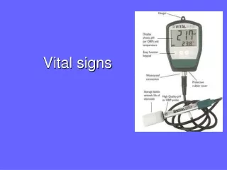
Vital Signs • Important indicators that provide information about the basic body conditions of the patient • Four main vital signs: temperature, pulse, respirations, and blood pressure
Temperature • Definition – a measurement of the balance between heat lost and heat produced by the body
Temperature • Heat is produced by the metabolism of food and muscle, and gland activity • Heat is lost through perspiration, respiration, and excretion • Homeostasis (constant state of fluid balance) is the ideal state in the human body • Normal temperature range is 97 to 100 degrees Fahrenheit or 36.1 to 37.8 degrees Celsius
Temperature • Aseptic technique for handling • Oral measurement – mouth; 3-5 min • Rectal measurement – rectum; 3-5 min • Axillary measurement – armpit; 10 min • Aural measurement – ear; less than 2 sec
Temperature • Hypothermia – very low body temperature, below 95 degrees Fahrenheit measured rectally • Factors that lead to decreased body temperature: starvation or fasting, decreased muscle activity, mouth breathing, exposure to cold environmental temperatures and certain diseases • Hyperthermia – body temperatures exceeds 104 degrees Fahrenheit measured rectally • Factors that lead to increased body temperature: illness, infection, exercise, excitement, and high environmental temperatures
Temperature • Usually measured on Fahrenheit scale, but may see Celsius scale • Convert Fahrenheit to Celsius temperature, subtract 32 from the Fahrenheit temperature and then multiply the result by 5/9 or 0.5556. • Convert Celsius to Fahrenheit temperature, multiply the Celsius temperature by 9/5 or 1.8 and then add 32 to the total.
Temperature • Equipment used for measurement • Clinical thermometer – slender glass tube containing mercury, which expands when exposed to heat • Electronic thermometer – usually battery operated unit registering temperature in about 60 sec on digital display screen • Tympanic thermometer – specialized electronic thermometer measuring temperature in the auditory canal in 1 to 2 sec on display screen
Temperature • Conditions requiring modifications • Oral - eating, drinking, or smoking • Rectal – diarrhea • Axillary or groin – moisture or rubbing • Aural – incorrect positioning of pinna Recording or documenting - T • Cleaning equipment
Pulse • Definition- the pressure of the blood felt against the wall of an artery as the heart contracts and relaxes, or beats • Felt in arteries that lie fairly close to the skin and can be pressed against a bone by the fingers
Pulse • Pulse sites • Temporal – at side of forehead • Carotid – at neck • Brachial – at inner aspect of forearm at antecubital space • Radial – at inner aspect of wrist above thumb; most common site • Femoral – at inner aspect of upper thigh • Popliteal – behind knee • Dorsalispedis – at top foot arch
Pulse • Measured in number of beats per minute • Rate – number of beats per minute • Rhythm – regularity; regular or irregular • Volume – strength; strong, weak, thready, or bounding
Pulse • Pulse rates vary depending on age, sex, and body size • Adults – 60 – 90 bpm • Men – 60 – 70 bpm • Women – 65 – 80 bpm • Children over 7 years – 70 – 90 bpm • Children 1 to 7 years – 80 – 100 bpm • Infants – 100 – 160 bpm
Pulse • Bradycardia – pulse under 60 bpm • Tachycardia – pulse over 100 bpm • Arrhythmia – irregular or abnormal rhythm
Pulse • Factors that may change pulse rate • Increased – exercise, stimulant drugs, excitement, fever, shock, and nervous tension • Decreased – sleep, depressant drugs, heart disease, coma, and physical training • Document findings - P
Respirations • Definition- the process of taking in oxygen and expelling carbon dioxide from the lungs and respiratory tract • One respiration consists of one inspiration and one expiration
Respirations • The breathing rate of the patient • Rate – number of respirations per minute = • Adults – 12 –20 rpm • Children – 16 – 25 rpm • Infants – 30 –50 rpm • Rhythm – regularity = regular or irregular • Character – type; depth and quality = deep, shallow, labored, difficult, stertorous, and moist
Respirations • Abnormal respirations • Dyspnea- difficult or labored breathing • Apnea – absence of breathing • Tachypnea – respiratory rate above 25 rpm • Bradypnea – respiratory rate below 10 rpm • Orthopnea – difficult breathing in any position other than sitting erect or standing
Respirations Abnormal respirations continued • Cheyne-Stokes respirations – periods of dyspnea followed by periods of apnea = frequently seen in dying patients • Rales – bubbling or noisy sounds caused by fluids or mucus in the air passages
Respirations • Must be counted in such a way that patient is unaware of the procedure as respirations are partially under voluntary control • Leave hand on pulse site while counting respirations • Document findings - R
Blood Pressure • Definition– force exerted by the heart against the arterial walls when the heart contracts or relaxes • Read in millimeters (mm) of mercury (Hg)
Blood Pressure • Two types of measurements • Systolic – pressure in the walls of the arteries when the heart is contracting and pushing blood into the arteries • Reading shows greatest pressure • Normal reading is 120 mm Hg • Normal range is 100 to 140 mm Hg • Diastolic – constant pressure in the walls of the arteries when the heart is at rest or between contractions • Reading shows least pressure • Normal reading is 80 mm Hg • Normal range is 60 to 90 mm Hg
Blood Pressure • Pulse pressure – difference between systolic and diastolic pressure • Important indicator of health and tone of arterial walls • Normal range in adults 30 to 50 mm Hg
Blood Pressure • Hypertension – high blood pressure • Systolic greater than 140 mm Hg • Diastolic greater than 90 mm Hg • Causes – stress, anxiety, obesity, high-salt intake, aging, kidney disease, thyroid deficiency and vascular conditions
Blood Pressure • Hypotension – low blood pressure • Systolic less than 100 mm Hg • Diastolic less than 60 mm Hg • Causes – heart failure, dehydration, depression, severe burns, hemorrhage, and shock
Blood Pressure • Factors influencing blood pressure readings • Force of heartbeat • Resistance of the arterial system • Elasticity of the arteries • Volume of the blood in the arteries
Blood Pressure • Factors increasing blood pressure • Excitement, anxiety, nervous tension • Stimulant drugs • Exercise and eating • Factors decreasing blood pressure • Rest or sleep • Depressant drugs • Shock • Excessive loss of blood • Factors causing miscellaneous readings • Lying down • Sitting position • Standing position
Blood Pressure • Recorded as a fraction • Systolic is numerator = top number • Diastolic is denominator = bottom number • Sphygmomanometer instrument used to measure blood pressure = B/P cuff • Mercury – long column; 2mm marks • Aneroid – face scale; 2mm marks • Parts: cuff, bladder, control valve, bulb, tubing, measurement scale • Stethoscope: earpieces, diaphragm, bell and tubing • Document findings – B/P
Apical Pulse • Definition– pulse count taken at the apex of the heart with a stethoscope • Stethoscope amplifies the actual heart beat • Usually ordered on patients with irregular heartbeats, hardening of the arteries, or weak or rapid radial pulses, and infants • Two separate heart sounds are heard while listening to the heartbeat = “lubb-dupp” caused by closing of heart valves as blood flows through chambers of the heart • Each lubb-dupp counts as ONE heartbeat
Apical Pulse • Pulse deficit – heart condition in which heart is weak and does not pump enough to blood to produce a pulse or heart beats too fast and there is not enough time for the heart to fill with blood • The heart does not produce a pulse during each beat • Apical pulse rate is higher than the pulse rate at the other pulse sites on the body • Most accurate determination of pulse deficit calculated by two persons at the same time • Document findings - AP
Documentation • Graphing vitals • Graphic sheets used for recording vitals • Visual diagram of variations in patient’s vital signs • Must be neat, legible, and accurate • Correct errors carefully