Download

1 / 18
190 likes | 330 Vues
PHYSIOLOGY LAB EEG I . The cerebral cortex is composed of nerve cells, many of which are functionally connected to each other, and connected to other parts of the brain.

E N D
The cerebral cortex is composed of nerve cells, many of which are functionally connected to each other, and connected to other parts of the brain. • Electrical activity in the form of nerve impulses being sent and received to and from cortical neurons is always present, even during sleep.
EEG • Since the cerebral cortex just under the cranium, electrodes placed on the scalp above the various regions of the brain can detect the electrical activity associated with functioning neurons. • The recording of the brain’s activity obtained by using electrodes is called “electroencephalography: EEG”
Usage • Epilepsy for diagnosis, localization, prognosis and control of treatment • Brain Trauma for aid in the diagnosis, localization and prognosis • Brain abscess for aid in the diagnosis and localization • Subdural hematoma for aid and localization • Cerebral hemorrhage for aid in the diagnosis and localization • Cerebral thrombosis for aid in the diagnosis and localization • Cerebral palsy for detection of epileptic disorder • Meningitis for detection of residuals • Encephalitis for diagnosis and prognosis • Behavior disorder for detection of possible epileptic disorder or organic brain disease • Narcolepsy for detection of possible epileptic disorder or organic brain disease • Migraine for detection of possible epileptic disorder or organic brain disease • Syncope for detection of possible epileptic disorder or organic brain disease • Intellectual defects for detection of possible epileptic disorder or organic brain disease • General surgery for control of anesthesia • Heart & lung surgery for monitoring the oxygen supply to the brain • Intensive care for determining whether the patient is recovering, sinking deeper into coma or if the patient is in a state of "irreversible coma" in which higher cognitive functions cannot be restored.
EEG Source • The pyramidal neurons extending perpendicular to the cortex is the main signal source of the EEG waves.
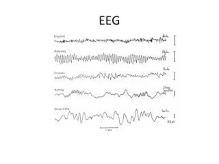
Alpha • In general the alpha rhythm is the prominent wave pattern of an adult who is awake but relaxed with closed eyes. • In general alpha waves are diminish when people open their eyes and are attentive to external stimuli.
Beta • Beta rhythm occurs when people are alert and attentive to external stimuli, or exert specific mental effort. • The amplitude of beta rhythm is lower than the alpha r. • Thus instead of getting the wave like synchronized pattern of alpha waves desynchronization or alpha block occurs
Delta and theta • These waves are low frequency EEG patterns that increase during sleep in the normal adult • As people move from lighter to deeper sleep stages, the alpha waves diminishes and is replaced by the lower freq. Theta and delta waves.
Gamma • Gamma rhtythms are high freq. EEG patterns that can be recorded from all lobes of the cerebrum.
The experiment • 1 positive electrod • 1 negative electrod • 1 ground-neutral electrod Electrode placement and lead attachment
Calibration Clench Fist for Calibration
Recording • Seconds 0-10 eyes closed • Seconds 10-20 eyes open • Seconds 20-30 eyes re-closed • Insert marker • at 10 sec eyes open • At 20 sec eyes re-closed • After the recording click on the freq. Buttons in the following sequence: • Alpha, beta, delta, theta
Data Analysis • std dev values are going to be analyzed. • St dev: is a measure of the variability of data points. The data represents amplitudes of the brain rhythms. The advantage of the st dev measurement is that extreme values or artifacts do not unduly influence the measurement.
Analysis Eyes open Eyes closed Eyes re-open