Download
1 / 25
250 likes | 444 Vues
From molecule to memory in the cerebellar neural circuit. Sang Jeong Kim Department of Physiology Seoul National University College of Medicine, Korea. Linden DJ (2003) Science 301. Cerebellar cortical circuit. Purkinje cell (PC) - Main sole output of cerebellar cortex. Sensory-motor

E N D
From molecule to memory in the cerebellar neural circuit Sang Jeong Kim Department of Physiology Seoul National University College of Medicine, Korea
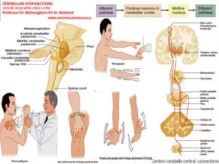
Linden DJ (2003) Science301. Cerebellar cortical circuit Purkinje cell (PC) - Main sole output of cerebellar cortex Sensory-motor input Error signals Motor output Two inputs to PC : parallel fiber (PF) climbing fiber (CF)
Pairing of PF and CF induces long-term depression (LTD) of PF-PC synapse Parallel fiber Pairing Tone 180 160 140 LTD 120 Purkinje Cell response 100 80 60 Climbing fiber Shock 40 -10 -5 0 5 10 15 20 25 30 35 Time (min) LTD = Memory trace
Eye-Blink Conditioning:A simple form of associative motor learning LTD = memory trace -> Tone means Shock Linden DJ (2003) Science301.
PF-PC synapse Presynaptic Terminal of PF • Excitatory glutamatergic synapse • Single firing of PF evokes fast excitatory postsynaptic current (fast EPSC) via AMPA receptor (GluR2-containing, Ca2+-impermeable variety). • Mature PC has no NMDA receptor. • Pairing of PF and CF induces long-term depression (LTD) which is internalization of AMPA-R in dendritic spines of PC. • AMPA-R LTD is a memory trace • PF drives PC up to 100 Hz. Glutamate Spine of PC AMPA-R
Metabotropic glutamate receptor type1 (mGluR1) Presynaptic Terminal of PF • Single firing of PF : fast EPSC only • Tetanic burst stimulation of PF • Spillover of glutamate to perisynaptic region • mGluR1 in the perisynaptic region of the spine • Activation of mGluR1 by burst only Glutamate Spine of PC AMPA-R mGluR1 LTD is mGluR1-dependent: mGluR1 knock-out mice show defects in PF-PC LTD and motor learning
mGluR1-mediated signaling in PF-PC synapse Presynaptic Terminal of PF • mGluR1 signal has two limbs • 1) PLC pathway • IP3-mediated Ca release • 2) Activation of membrane conductance • Slow excitatory postsynaptic current (slow EPSC) Glutamate Spine of PC AMPA-R DAG Gq Ca2+ IP3 PLCb4 mGluR1 pump Ca2+ TRPC1 IP3R1 Na+, Ca2+ Ca2+ RyR Endoplasmic reticulum
Tetanic stimulus to PF induces mGluR1-evoked slow EPSC in Purkinje cells 100 pA 0.5 s 2,5,10,15 pulses in 100 Hz, 14 mA tetanus at -70 mV 10 mM CNQX 04020318-22
The slow EPSC evoked by parallel fiber bursts is mediated by an mGluR1-TRPC1 pathway Slow EPSC is also blocked by a dominant-negative TRPC1 and a TRPC1 Ab Kim et al, 2003 nature
LTD of the parallel fiber-evoked mGluR1-mediated slow EPSC by strong depolarization Pre PF burst (10 pulses, 100 Hz)
LTD of the parallel fiber-evoked mGluR1-mediated slow EPSC by strong depolarization Pre 30 s after depol PF burst (10 pulses, 100 Hz)
depolarization LTD of the parallel fiber-evoked mGluR1-mediated slow EPSC by strong depolarization A Pre 30 s after depol 1200 s after depol n=11 120 100 80 Current amplitude (%) 60 40 200 pA 0.5 s ** 20 PF burst (10 pulses, 100 Hz) 0 Fast EPSC( ) Slow EPSC( ) B 120 1 s n=14 n=11 100 2 s 100 * 5 s 80 80 60 Slow EPSC (%) 60 Slow EPSC (%) 40 40 n=11 20 20 ** 0 0 Time (s) 1 2 5 -300 0 300 600 900 1200 1500 Duration of depolarization (s)
200 pA 1 s LTD(mGluR1) is blocked by removing external Ca depolarization (n=14) A Ca-free 120 90 s after depolarization Pre 100 80 Current amplitude (%) 60 Fast current Slow current DHPG/Glu 40 20 Time (min) -6 -4 -2 0 2 4 6 B depolarization (n=11) Ca-free 120 Pre Ca-free ACSF 5 min after depolarization 100 80 Current amplitude (%) 60 40 Fast EPSC Fast EPSC 200 pA Slow EPSC Slow EPSC PF burst 20 0.5 s 0 Time (min) -10 -5 0 5 10 15 20
A dynamin blocker, dynasore inhibited LTD of mGluR1 Before depolarization 30 min after depolarization
What is the function of LTD of mGluR1 in physiological and patho-physiological conditions?
Physiological CF bursts produce LTD of mGluR1-mediated dendritic Ca transients Pre-drug +CPCCOEt 100 % 1 s 1 mm PF burst CF burst x 50 50 % 20 s dF/F 1 nA CF burst x 50 100 % 1st burst 2nd burst 50th burst 1 s 20 mV 20 ms 1.2 s
LTD(mGluR1) blocks subsequent LTD of AMPA-Rs B After LTD(mGluR1) (n=8) Control (n=9) A 100 pA 100 pA 0.1 s 0.1 s pairing pairing 180 180 160 160 140 140 120 120 Normalized EPSC (%) Normalized EPSC (%) 100 100 80 80 60 60 40 40 Time (min) Time (min) -10 -5 0 5 10 15 20 25 30 35 -10 -5 0 5 10 15 20 25 30 35 C PF 5 stim. @ 100 Hz X 30, 2 sec interval PC 0 mV for 75 ms
120 100 80 60 40 20 0 600 48hr 6hr con 24hr 1hr 500 120 400 100 % of PI uptake 300 80 60 200 40 100 20 0 0 48hr con 24hr 6hr 1hr Control OGD(100min) LTD of mGluR1 in a Ischemia Model: Oxygen-Glucose Deprivation in Organotypic Slice Culture 48hr after OGD 24hr after OGD 6hr after OGD 1hr after OGD Surface expression of mGluR1a Con OGD(100min) Control Surface mGluR1a Total mGluR1a DIC Actin Total expression of mGluR1a PI staining
Internalization of mGluR1 as expression mechanism of LTD of mGluR1 Spine Internalization ?? mGluR1 Depolarization CF Hypoxia mGluR1 LTD of mGluR1 AMPA 수용체 (AMPAR)
Taking advantages of multiphoton-microscopy • Higher axial resolution • Monitoring of tagged mGluR1 distribution • Greater sample penetration • Dendritic and axonal mobility in the intact brain such as spinal cord Multi-photon Single-photon OGB-1, Zeiss LSM 510
Implementation mGluR1/TRPC signaling Identification Coincidence detector for AMPA-R LTD Function Regulation LTD of mGluR1/TRPC Control Mechanisms of mGluR1 internalization Tool Development Peptide delivery to Purkinje cell Vestibulo-ocular reflex Apply to Behavior
Acknowledgements Lab of Neuronal information storage, Seoul National University College of Medicine (http://brain.snu.ac.kr) • Sang Jeong Kim • Jun Kim • Hong Goo Chae • Yunju Jin • Yon Wha Hong • Hae Young Kim • Lyan Choi • Won Sok Chang • Sung Soo Chang • Ji Young Kim • Sung Won Hur • Chang Hee Kim Johns Hopkins University: David Linden Lab, Paul Worley Lab