Download
1 / 106
1.1k likes | 1.65k Vues
Chapter 17: Blood. Overview. Blood functions Compostion of whole blood Plasma RBCs – structure, function, and development Blood types WBCs Platelets Hemostasis. The Cardiovascular System. A circulating transport system composed of: a pump (the heart)

E N D
Overview • Blood functions • Compostion of whole blood • Plasma • RBCs – structure, function, and development • Blood types • WBCs • Platelets • Hemostasis
The Cardiovascular System • A circulating transport system composed of: • a pump (the heart) • a conducting system (blood vessels) • a fluid medium (blood) • Functions to transport: • oxygen and carbon dioxide • nutrients • hormones • immune system components • waste products
General Characteristics of Blood • Blood is a sticky, opaque fluid with a metallic taste • Color varies from scarlet to dark red High viscosity (due to cells) • Temperature is 38C • Normal pH range = 7.35–7.45 • Blood volume (liters) = 7% of body weight (kilograms): • adult male: 5 to 6 liters • adult female: 4 to 5 liters
Blood - General Functions • Transport of dissolved gases, nutrients, hormones, and metabolic wastes • Regulation of pH, body temperature, ion composition of interstitial fluids • Restriction of fluid loss at the injury site • Defense against toxins and pathogens
Whole Blood • Plasma: Fluid component • Water (90%) • Dissolved plasma proteins • Other solutes • Formed elements: Cells and fragments • RBCs (carry Oxygen) • WBCs (immunity) • Platelets (cell fragments involved in clotting)
Plasma Figure 19–1b
Plasma • Makes up 50–60% of blood volume • More than 90% of plasma is water • Other constiuents: • Plasma proteins • Lactic acid, urea, creatinine • Organic nutrients – glucose, carbohydrates, amino acids • Electrolytes – sodium, potassium, calcium, chloride, bicarbonate • Respiratory gases – oxygen and carbon dioxide
Body Fluids Extracellular Fluid (ECF) = Interstitial fluid (IF) and plasma plus a few other body fluids such as CSF • Plasma and IF exchange water, ions, & small solutes across capillary walls Intracellular Fluid (ICF)=fluid inside cells ECF and ICF differ in their levels of: • O2 and CO2 • Dissolved proteins: plasma proteins do not pass through capillary walls (too large)
Plasma proteins • Albumins (60%): major component of osmotic pressure of plasma • Transport proteins for fatty acids, thyroid hormones, steroid hormones • Globulins (35%): antibodies (immunoglobulins) and transport proteins: • hormone-binding proteins • metalloproteins • apolipoproteins (lipoproteins) • steroid-binding proteins • Fibrinogens (4%)-functions in blood clotting (form fibrin) • Others (1%) including hormones
Origins of Plasma Proteins • 90% made in liver • Others not made in the liver include: • Antibodies made by plasma cells (a special type of WBC) • Peptide hormones made by endocrine organs
Serum • Liquid part of a blood sample in which dissolved fibrinogen has converted to solid fibrin • Often, this term refers to plasma that has had the clotting proteins removed
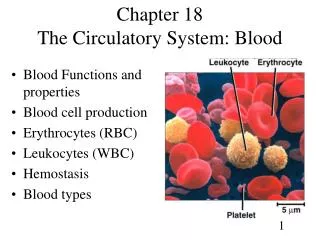
Formed Elements • These are the cells (and quasi-cellular) constituents of blood • Red blood cells (RBCs) make up 99.9% of blood’s formed elements • White blood cells and platelets make up the rest
Components of Whole Blood Figure 17.2
Measuring RBCs • Red blood cell count: reports the number of RBCs in 1 microliter whole blood • Male: 4.5–6.3 million • female: 4.2–5.5 million • Hematocrit (packed cell volume, PCV): percentage of RBCs in centrifuged whole blood • male: 40–54 (avg = 46) • female: 37–47 (avg = 42) RBCs make up about 1/3 of all cells in the body!
Why do RBCs look hollow? No nucleus Biconcave structure
RBC Structure • Small and highly specialized disc • Thin in middle and thicker at edge Why this structure? Figure 19–2d
Importance of RBC Shape and Size • High surface-to-volume ratio: • Increase surface area for gas exchange • Discs form stacks: • smoothes flow through narrow blood vessels • Discs bend and flex entering small capillaries: • 7.8 µm RBC passes through 4 µm capillary
Shaped like biconcave discs Function primarily to carry oxygen-contain hemoglobin (95% of RBC protein) Lack a nucleus and contain few organelles (no mitochondria, ribosomes) Life span approx. 120 days Generate ATP anaerobically (no mitochondria) so they don’t consume any of the oxygen that they transport RBC characteristics
Hemoglobin(Hb) • Protein molecule inside RBCs that transports respiratory gases • Composed of: • Fourprotein chains called globins • adults: 2 alpha and 2 beta chains • Each of these four chains is bound to a pigment molecules called heme • each of which contain one iron ion (red color) and bind one oxygen molecule • Each RBC ~280 million molecules
Hemoglobin Structure • Complex quaternary structure Figure 19–3
Fetal Hemoglobin (Hb F) • Made up of 2 alpha and 2 gamma chains • Has a higher affinity for oxygen than adult hemoglobin, “steals” oxygen from maternal hemoglobin in utero
RBC fate After 100-120 days: • 10% hemolyze in the blood • 90% removed by macrophages in the spleen (especially), the liver and the bone marrow and heme is recycled: • heme degraded to biliverdin (green) • biliverdin converted to bilirubin (yellowish) • Bilirubin leaves Mphage, binds to albumin, tranported to liver for excretion in bile (high levels of bilirubin in jaundice) • In colon, bacteria convert bilirubin to urobilinogens and stercobilinogens – colors feces • Some is absorbed into circulation and eliminated by kidneys in urine – colors urine
Serum Bilirubin • Red cells account for 85% of bilirubin formed = Unconjugated • In liver it is conjugated and secreted into bile to large intestine • Hemolytic jaundice: elevated levels of unconjugated bilirubin • Obstructive jaundice: elevated levels of conjugated bilirubin because bile ducts are blocked (bile that can’t be secreted)
Recycling • Iron • Heme iron is removed in spleen (or liver or bone marrow) • Binds to plasma protein called transferrin • Transferrin is taken up in bone marrow and used to make new heme in developing RBCs • Very efficient • Globin protein • Amino acids travel through bloodstream to bone marrow and can be used in erythropoiesis
Hematopoiesis • Development of all the cells of the lymphoid/myeloid lineage • Includes: RBCs, all types of WBCs, and platelets • All start out as hemocytoblasts, a pluripotent stem cell: • Myeloid stem cells give rise to RBCs, platelets and some WBCs • Lymphoid stem cells give rise to lymphocytes only • Occurs in red bone marrow (axial and epiphyses)
Erythropoiesis • Rate of RBC production controlled by erythropoietin - EPO (from where?) • What is necessary for healthy RBCs? • amino acids • iron • vitamins B12, B6, and folic acid
Erythropoietin Mechanism Imbalance Start Homeostasis: Normal blood oxygen levels Stimulus: Hypoxia due to decreased RBC count, decreased amount of hemoglobin, or decreased availability of O2 Imbalance Increases O2-carrying ability of blood Reduces O2 levels in blood Kidney (and liver to a smaller extent) releases erythropoietin Enhanced erythropoiesis increases RBC count Erythropoietin stimulates red bone marrow Figure 17.6
RBC Maturation • Hematocytoblast myeloid stem cell proerythroblast erythroblast reticulocyte mature RBC • Reticulocytes have no nucleus and enter bloodstream still containing ribosomes and mRNA. After a day or so of furious Hb production, lose their organelles and become mature RBCs Figure 19–5
Regulation of Erythropoiesis • Circulating erythrocytes – the number remains constant and reflects a balance between RBC production and destruction • Too few RBCs leads to tissue hypoxia • Too many RBCs causes undesirable blood viscosity
Blood Types • Genetically determined cell surface markers (antigens) on RBCs, including • ABO group – glycolipids on RBC surface • Rh factor – membrane protein
4 Basic Blood Types • A has surface antigen A • B has surface antigen B • AB has both antigens A and B • O has neither A nor B • A has type B antibodies • B has type A antibodies • O has both A and B antibodies • AB has neither A nor B antibodies
4 Basic Blood Types • Antigens also called agglutinogens • Antibodies called agglutinins Figure 19–6a
The Rh Factor • Also called D antigen • Either Rh positive (Rh+) or Rh negative (Rh-) • Only sensitized Rh- blood has anti-Rh antibodies
Cross-Reaction Figure 19–6b
Cross-Reaction • If donor and recipient blood types not compatible: • Plasma antibody meets its specific surface antigen and blood will agglutinate and hemolyze
Blood Type Test • Determines blood type and compatibility Figure 19–7
Cross-Match Test • Performed on donor and recipient blood for compatibility to blood surface antigens other than ABO and Rh
Blood type questions • Which blood type is the best in emergency settings (hint: which type can be given to anyone?) • Which blood type is the lucky one that can receive blood from any donor?
Hemolytic Disease of the Newborn (Erythroblastosis Fetalis) • Mother is Rh- • Father and fetus are Rh+ • First pregnancy = sensitization at delivery due to hemorrhage • Second pregnancy = Anti-Rh IgG antibodies can cross placenta to attack fetal RBCs hemolysis and excess presence of erythroblasts
Second pregnancy is attacked by maternal antibodies Treatment?
Transfusions • Unit whole blood = 500ml • About half of this is plasma which contains antibodies. There is a slight risk of graft versus host (GVH) reactions, but since the volume in one unit is only about 10% of total plasma volume, usually gets diluted out • If RBCs are needed, can use packed RBCs instead of whole blood
White Blood Cells (WBCs) • Leukocytes: the only blood components that are complete cells; have nuclei and other organelles, not involved in oxygen transport. • Functions: • Defend against pathogens • Remove toxins and wastes • Attack abnormal cells