Download
1 / 49
1k likes | 2.52k Vues
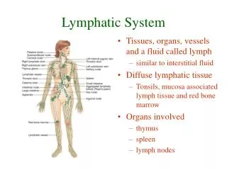
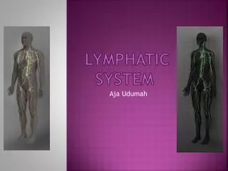
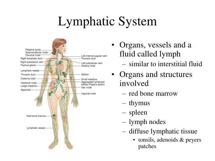
Lymphatic System. Organs, vessels and a fluid called lymph similar to interstitial fluid Organs and structures involved red bone marrow thymus spleen lymph nodes diffuse lymphatic tissue tonsils, adenoids & peyers patches. Functions of the Lymphatic System.

E N D
Lymphatic System • Organs, vessels and a fluid called lymph • similar to interstitial fluid • Organs and structures involved • red bone marrow • thymus • spleen • lymph nodes • diffuse lymphatic tissue • tonsils, adenoids & peyers patches
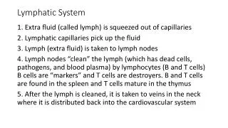
Functions of the Lymphatic System • Draining excess interstitial fluid from tissue spaces • Transporting dietary lipids & vitamins from GI tract to the blood • Facilitating immune responses
Lymphatic Vessels & Circulation • Capillaries that begin asclosed-ended tubes foundin spaces between cells • Combine to form lymphaticvessels • resemble veins with thinwalls & more valves • Fluid flows through lymph nodes towards large veins (subclavian veins) above the heart • lymph emptied into bloodstream
Lymphatic Capillaries • Found throughout thebody except in Avasculartissue (cartilage, epidermis& cornea) • Structure is designed to lettissue fluid in but not out
Formation & Flow of Lymph • Fluid & proteins escaping from vascular capillaries is collected by lymphatic capillaries & returned to the blood • Lymphatic vessels empty into subclavian veins
Lymphatic Organs & Tissues • Widely distributed throughout the body • Primary lymphatic organs • provide environment for stem cells to divide & mature into B and T lymphocytes • red bone marrow gives rise to mature B cells • thymus is site where T cells mature • Secondary lymphatic organs & tissues • site where most immune responses occur • lymph nodes, spleen & lymphatic nodules
Thymus Gland (Primary lymphatic organ) • Large organ in infants (70 g) but atrophied as adult (3 g) • 2 lobed organ located in mediastinum • Each lobule has cortex &medulla • Cortex • tightly packed lymphocytes,macrophages, and epithelial cells • Epithelial cells help “educate” T cells • Medulla • Same cells but less dense • Hassall’s corpuscles- clusters of dying cells, function unknown
Lymph Nodes (secondary lymphatic organ)
Lymph Nodes • Bean-shaped organs, up to 1 inch long, located along lymphatic vessels • scattered throughout body but concentrated near mammary glands, axillae & groin • cortex • lymphatic nodules containing dendritic cells • antigen-presenting cells and macrophages • B cells proliferate into antibody-secreting plasma cells • medulla • contains B cells & plasma cells in a network of reticular fibers and reticular epithelial cells
Lymph Nodes • Flow is in one direction • afferent vessels lead in • sinuses lead to efferent vessels that exit at hilus • Only nodes filter lymph
Metastasis Through Lymphatic System • Characteristic of malignant tumors • Spread of disease from one organ to another • cancer cells travel via blood or lymphatic system • cells establish new tumors where they lodge • Secondary tumor sites can be predicted by direction of lymphatic flow from primary site • Cancerous lymph nodes are firm, enlarged and nontender -- infected lymph nodes are not firm and are very tender
Spleen-secondary lymphatic organ • 5 inch organ between stomach & diaphragm • Hilus contains blood & lymphatic vessels • White pulp and red pulp • white is lymphatic tissue (lymphocytes & macrophages) around branches of splenic artery • red pulp is venous sinuses filled with blood & splenic tissue (splenic cords)
Functions of Spleen White pulp: Lymphocytes and macrophages destroy foreign substances Red pulp: Removal of damaged blood cells Storage of platelets Production of blood cells during fetal life
Lymphatic Nodules • Concentrations of lymphatic tissue not surrounded by a capsule scattered throughout connective tissue of mucous membranes • mucosa-associated lymphoid tissue (MALT) • Peyer’s patches in the ileum of the small intestine • Appendix • Tonsils form ring at top of throat • adenoids (pharyngeal tonsil) • palatine tonsils (on each side wall) • lingual tonsil in the back of the tongue
Resistance Ability to ward of damage or disease • Nonspecific resistance • general defensive mechanisms effective on a wide range of pathogens • Specific resistance (immunity) • Ability to fight a specific pathogen • cell-mediated immunity (T cells) • antibody-mediated immunity (B cells)
Nonspecific Resistance to Disease • Immediate protection against wide variety of pathogens & foreign substances • lacks specific responses to specific invaders • Mechanisms function regardless of type of invader • external mechanical & chemical barriers • internal nonspecific defenses • antimicrobial proteins • natural killer cells & phagocytes • inflammation & fever
Skin & Mucous Membranes • Mechanical protection • skin (epidermis) closely packed, keratinized cells • shedding helps remove microbes • mucous membrane secretes viscous mucous • cilia & mucus trap & move microbes toward throat • washing action of tears, urine and saliva • Chemical protection • sebum inhibits growth bacteria & fungus • perspiration lysozymes breakdown bacterial cells • acidic pH of gastric juice and vaginal secretions destroys bacteria
Internal Defenses • Antimicrobial proteins discourage microbial growth • interferons • produced by virally infected lymphocytes & macrophages • diffuse to neighboring cells to induce synthesis of antiviral proteins • complement proteins • inactive proteins in blood plasma • when activated enhance immune, allergic & inflammatory reactions • transferrins • iron-binding proteins inhibit bacterial growth by reducing available iron
Natural Killer Cells & Phagocytes • NK cells kill a variety of microbes & tumor cells • found in blood, spleen, lymph nodes & red marrow • attack cells displaying abnormal MHC antigens • Phagocytes (neutrophils & macrophages) • ingest microbes or particulate matter • macrophages developed from monocytes • fixed macrophages stand guard in specific tissues • kupffer cells in the liver • wandering macrophages in most tissue
Phagocytosis • Chemotaxis • attraction to chemicals from damaged tissues, complement proteins, or microbial products • Adherence • attachment to plasma membrane of phagocyte • Ingestion • engulf by pseudopods to form phagosome • Digestion & killing • merge with lysosome containing digestive enzymes • exocytosis residual body
Inflammation • Damaged cell initiates • Signs of inflammation • redness • heat • swelling • pain • Function is to trap microbes, toxins or foreign material & begin tissue repair
Fever • Abnormally high body temperature that occurs because the hypothalamic thermostat is reset • Occurs during infection & inflammation • bacterial toxins trigger release of fever-causing cytokines such as interleukin-1 • Benefits • intensifies effects of interferons, inhibits bacterial growth, speeds up tissue repair
Specific Resistance: Immunity • Immunity is the bodies ability to defend itself against specific foreign material or organisms • bacteria, toxins, viruses, cat dander, etc. • Differs from nonspecific defense mechanisms • specificity----recognize self & non-self • memory----2nd encounter produces even more vigorous response • Immune system is cells and tissues that produce the immune response • Immunology is the study of those responses
Maturation of T and B Cells • T cell mature in thymus • cell-mediated response • Cell directly attacks the invading antigen • effective against fungi, viruses, parasites, cancer, and tissue transplants • B cells in bone marrow • antibody-mediated response • plasma cells secrete antibodies which affect antigens • effective against bacteria
Antigens • Molecules or bits of foreign material • entire microbes, parts of microbes, bacterial toxins, pollen, transplanted organs, incompatible blood cells • Required characteristics to be considered an antigen • immunogenicity = ability to provoke immune response • reactivity = ability to react to cells or antibodies • Get past the bodies nonspecific defenses • enter the bloodstream to be deposited in spleen • penetrate the skin & end up in lymph nodes • penetrate mucous membrane & lodge in associated lymphoid tissue
Chemical Nature of Antigens/Epitopes • Large, complex molecules, usually proteins • if have simple repeating subunits are not usually antigenic (plastics in joint replacements) • small part of antigen that triggersthe immune response is epitope Antigen
Diversity of Antigen Receptors • Immune system can recognize and respond to a billion different epitopes -- even artificially made molecules • Explanation for great diversity of receptors is genetic recombination of few hundred small gene segments • Each B or T cell has its own unique set of gene segments that codes its unique antigen receptor in the cell membrane
Major Histocompatibility Complex Antigens • All our cells have unique surface markers (1000s molecules) • MHC-I molecules are found in cell membrane of all cells except red blood cells • MHC-II markers seen only on membrane of antigen presenting cells (macrophages, B cells, thymus cells) • Function • if cell is infected with virus MHC-I contain bits of virus marking cell so T cells recognize there is a problem • if antigen presenting cells (macrophages or B cells) ingest foreign proteins, they will display as part of their MHC-II
Pathways of Antigen Processing • B and T cells must recognize a foreign antigen before beginning their immune response • B cells can bind to antigen in extracellular fluid • T cells can only recognize fragments of antigens that have been processed and presented to them as part of a MHC molecule • Helper T cells “see” antigens if they are part of MHC-II molecules on surface of antigen presenting cell • Cytotoxic T cells “see” antigens if they are part of MHC-I molecules on surface of body cells
Processing of Exogenous Antigens • Foreign antigen in body fluid is phagocytized by APC • macrophage, B cell, dendritic cell (Langerhans cell in skin) • Antigen is digested and fragments are bound to MHC-II molecules stuck into antigen presenting cell membrane • APC migrates to lymphatic tissue to find T cells
Processing of Endogenous Antigens • Endogenous antigens are foreign proteins produced within a body cell --- viral or cancerous • Fragments of proteins become part of MHC-I molecules displayed at surface of cell • T cells recognize the antigen presented by the MHC-I molecule as foreign and initiates immune response.
Cell-Mediated Immunity • Begins with activation of T cell by a specific antigen • Result is T cell capable of an immune attack • elimination of the intruder by a direct attack
Activation, Proliferation & Differentiation of Cytotoxic T Cells • Receptor on T cell binds to foreign antigen fragment part of MHC-I • Costimulation from helper T cell • prevents accidental immune response • Proliferates & differentiates into population (clone) of Tc cells and memory Tc cells • Occurs in secondary lymphatic organs such as lymph node
Activation, Proliferation & Differentiation of Helper T Cells • Receptor on CD4 cell binds to foreign antigen fragment associated with MHC-II • Costimulation • Proliferates & differentiates into population (clone) of TH cells and long-lived memory TH cells
Types of Mature T Cells • Helper T cells (CD4) • Cytotoxic (killer) T cells (CD 8) • Memory T cells
Helper T Cells • Display CD4 on surface so also known as T4 cells or TH cells • Recognize antigen fragments associated with MHC-II molecules & activated by APCs • Function is to costimulate all other lymphocytes • secrete cytokines (small protein hormones) • autocrine function in that it costimulates itself to proliferate and secrete more interleukin (positive feedback effect causes formation of many more helper T cells)
Cytotoxic T Cells • Display CD8 on surface • Known as T8 or Tc or killer T cells • Recognize antigen fragments associated with MHC-I molecules • cells infected with virus • tumor cells • tissue transplants • Requires costimulation by cytokine from helper T cell
Memory T Cells • T cells from a clone that did not turn into cytotoxic T cells during a cell-mediated response • Available for swift response if a 2nd exposure should occur
Elimination of Invaders • Cytotoxic T cells migrate to site of infection or tumor formation • Recognize, attach & attack • secrete granules containing perforin that punch holes in target cell • secrete lymphotoxin that activates enzymes in the target cell causing its DNA to fragment • secrete gamma-interferon to activate phagocytic cells
Immunological Surveillance • Cancerous cell displays weird surface antigens (tumor antigens) • Surveillance = immune system finds, recognizes & destroys cells with tumor antigens • done by cytotoxic T cells, macrophages & natural killer cells • most effective in finding tumors caused by viruses • Transplant patients taking immunosuppressive drugs suffer most from viral-induced cancers
Antibody-Mediated Immunity • Millions of different B cells that can recognize different antigens and respond • B cells sit still and let antigens be brought to them • stay put in lymph nodes, spleen or peyer’s patches • Once activated, differentiate into plasma cells that secrete antibodies • Antibodies circulate in lymph and blood • combines with epitope on antigen similarly to key fits a specific lock
Activation, Proliferation, & Differentiation of B Cells • B cell receptors bind to antigen • Helper T cell costimulates • Rapid cell division & differentiation occurs • long-lived memory cells • clone of plasma cells • produce antibody at 2000 molecules/sec for 4-5 days • secrete only one kind antibody • Antibody enters the circulation to attack antigen
Antibody Structure • Glycoproteins called immunoglobulins • 4 polypeptide chains -- 2 heavy & 2 light chains • hinged midregion lets assume T or Y shape • tips are variable regions -- rest is constant region • 5 different classes based on constant region • IgG, IgA, IgM, IgD and IgE • tips form antigen binding sites
Antibody Actions • Neutralization of antigen by blocking effects of toxins or preventing its attachment to body cells • Immobilize bacteria by attacking cilia/flagella • Agglutinate & precipitate antigens by cross-linking them causing clumping & precipitation • Complement activation • Enhances phagocytosis
Role of the Complement System • Defensive system of plasma proteins that attack and destroy microbes • System activated by 2 different pathways • Produce same result • inflammation: dilation of arterioles, release of histamine & increased permeability of capillaries • opsonization: protein binds to microbe making it easier to phagocytize • cytolysis: a complex of several proteins can form holes in microbe membranes causing leakiness and cell rupture
Immunological Memory • Primary immune response • first exposure to antigenresponse is steady, slow • memory cells may remain fordecades • Secondary immune responsewith 2nd exposure • 1000’s of memory cells proliferate & differentiate into plasma cells & cytotoxic T cells • recognition & removal occurs quickly
Self-Recognition & Immunological Tolerance • T cells must learn to recognize self & lack reactivity to self proteins • T cells mature in thymus • those that can’t recognize self or react to it • destroyed by programmed cell death (apoptosis or deletion) • inactivated (anergy) -- alive but unresponsive • only 1 in 100 emerges immunocompetent T cell • B cells develop in bone marrow same way