Download
1 / 18
180 likes | 325 Vues
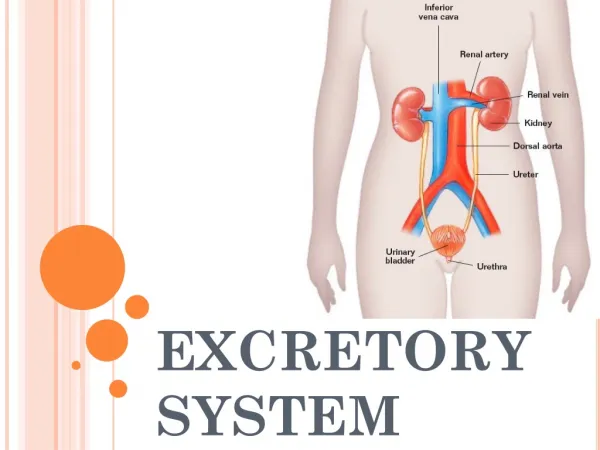
EXCRETORY SYSTEM. Adrenal glands. THE PARTS…. 2 kidneys 2 ureters 1 urinary bladder 1 urethra. 0. The KIDNEY. Fig. 18-2. Renal Cortex. Renal Medulla. Renal Artery. Renal Vein. Ureter. Adipose tissue/fat. 0. KIDNEYS ( INTERNAL ANATOMY ). The NEPHRON ….

E N D
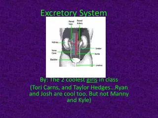
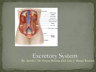
Adrenal glands THE PARTS… 2 kidneys 2 ureters 1 urinary bladder 1 urethra
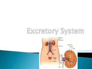
0 The KIDNEY Fig. 18-2 Renal Cortex Renal Medulla Renal Artery Renal Vein Ureter Adipose tissue/fat
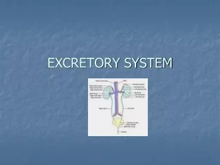
0 KIDNEYS(INTERNAL ANATOMY) The NEPHRON… • Functioning unit of the kidney • ~ 1 million / kidney • 2 main parts • Renal Corpuscle – expanded bulb-like end • Bowman’s capsule – bowl shaped structure that partially surrounds the capillary network called the… • Glomerulus – capillary network w/in the Bowman’s capsule that resembles a ball of yarn • Membrane is more permeable than most capillaries • High pressure
KIDNEYS(INTERNAL ANATOMY) • Renal tubule – thin twisted tube • Proximal convoluted tubule (PCT) – • Coiled segment that leads from its union (i.e. closest) w/Bowman’s capsule and then makes several turns • Loop of Henle – • Part of the tube that descends into medulla, makes a sharp U-turn, then ascends back toward the cortex. • Descending loop – descends from the PCT into the U-turn w/in the medulla • Ascending loop – ascends from the U-turn w/in medulla back into the cortex • Distal convoluted tubule (DCT) – • Coiled segment away from Bowman’s capsule that enters the collecting duct
KIDNEYS(INTERNAL ANATOMY) • Extra Parts • Collecting ducts – • Collect newly formed urine from DCT and channels it to the… ureter • Peritubular capillaries – • Porous, low-pressure capillaries that permit the exchange/movement of materials between the bloodstream and renal tubules
Blood Flow through the Nephron Pg. 537 Fig. 18-6 Renal corpuscle L H • Anatomy – difference in diameter between the afferent & efferent arterioles • Physiology – filtration • Boyle’s Law • 2. Anatomy – Glomerulus is more permeable than most capillaries (i.e. large pores) • Physiology – increase filtration • Hydrostatic pressure of the capillary (CAP-HP) Afferent arteriole Glomerulus Bowman’s Capsule filtrate Efferent arteriole PCT • Volume issues… • (45 gal/180 liters filtered)/day • do we “pee” this much? • so what must happen to it?
Capillary Exchange (pg. 387) Arteriole end Venous end • High CAP-HP • BP of capillaries • close to the heart • more fluid • Low CAP-HP • BP of capillaries • far from the heart • less fluid albumins • Hypertonic environment • ↑ solute • ↓ H2O plasma ISF • B) Hypotonic environment • ↓ solute • ↑H2O filtration reabsorption • ↑ CAP-COP - ↑ Capillary Colloidal Osmotic Pressure • albumins • less H2O • 4 Fluids • Plasma – blood volume • ISF – Interstitial fluid surrounding cells • Lymph – lymphatic vessels • CSF – cerebral spinal fluid
Capillary Colloidal Osmotic Pressure Plasma Proteins (pg. 347) • 3 main groups, primarily made by the liver and found in the blood • albumins • ~ 55% of the plasma proteins • Responsible for blood viscosity • Maintains bloods osmotic pressure • globulins • ~ 38% of the plasma proteins • Primarily help w/bodies immune response as antibodies • Fibrinogen • ~ 7% of the plasma proteins • Blood-clotting factor
Water and solutes from plasma pass from blood in glomerulus into Bowman’s capsule which empties into renal tubule. 1. Glomerular Filtration: • Pressure forces water and smaller solutes through the capillary membrane • capillaries are designed to allow small things through but not larger molecules, such as… • proteins, blood cells, and platelets (which should not be found in urine). • Glomerulus: specialized for its job • large surface area for filtration • thin, pourous membrane makes for a very “leaky” capillary • high capillary blood pressure (high pressure = more filtrate)
2. Tubular reabsorption returning of most of the filtered water and many filtered solutes to the blood stream • results in concentration of waste that remains in tubes to become urine • filtrate returns to blood • renal tubes peritubular capillaries (blood) • via active & passive (osmosis & diffusion) transport L • Tubular secretion movement of materials from blood and tubule cells into tubular fluid (H+, K+, ammonium …H+ controls blood pH) • Removal of harmful wastes not removed by filtration (reverse of reabsorption) • peritubular capillaries (blood) renal tubules • via active & passive (O/D) transport • 4. Anything that remains (metabolic waste) in the collecting duct eventually ends up in post nephron structures & secreted as urine H
0 KIDNEYS(INTERNAL ANATOMY) 3 tasks of nephron and collecting tubule: 1. Glomerular filtration 2. Tubular reabsorption 3. Tubular secretion 1. Glomerular Filtration: Water and solutes from plasma pass from blood in glomerulus into Bowman’s capsule which empties into renal tubule. • 150-180 liters/day • 99% returns back into blood • 1-2 liters is excreted as urine
0 KIDNEYS(INTERNAL ANATOMY) Pressure forces water and smaller solutes through the capillary membrane. Capillaries are designed to allow small things through but not larger molecules such as proteins, blood cells, and platelets (which should not be found in urine). 1. Glomerulus: specialized for its job (26.9) • large surface area for filtration • Thin, pourous membrane makes for a very “leaky” capillary • High capillary blood pressure (high pressure = more filtrate)
0 KIDNEYS(INTERNAL ANATOMY) 2. Tubular reabsorption returning of most of the filtered water and many filtered solutes to the blood stream • reabsorbed substances include: glucose, Na+, K+, Ca2+, Cl-, etc… 3. Tubular secretion movement of materials from blood and tubule cells into tubular fluid (H+, K+, ammonium …H+ controls the blood pH) Anything that remains in the collecting duct eventually ends up in renal pelvis and is secreted as urine Secretion in nephron results in formation of urine which is excreted from the body
0 KIDNEYS Fluid homeostasis in the body is controlled by the kidneys The hormone ADH (antidiuretic hormone) controls water reabsorption Fluid intake… • if high large volume of dilute urine • if low (or loss of fluid) small volume of concentrated urine Presence of ADH – more water reabsorbed = concentrated urine Absence of ADH – less water reabsorbed = dilute urine Alcohol inhibits secretion of ADH THE END