Download
1 / 22
380 likes | 835 Vues
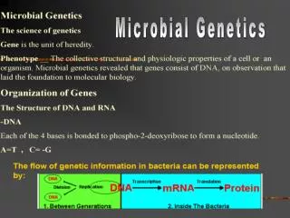
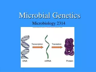
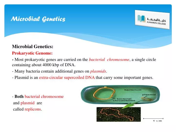
Microbial Genetics. Microbial Genetics : Prokaryotic Genome: - Most prokaryotic genes are carried on the bacterial chromosome , a single circle containing about 4000 kbp of DNA. - Many bacteria contain additional genes on plasmids .

E N D
Microbial Genetics Microbial Genetics: Prokaryotic Genome: - Most prokaryotic genes are carried on the bacterial chromosome, a single circle containing about 4000 kbp of DNA. - Many bacteria contain additional genes on plasmids. - Plasmid is an extra-circular supercoiled DNA that carry some important genes. - Bothbacterial chromosome and plasmid are called replicons.
Microbial Genetics - Genes essential for bacterial growth are carried on chromosome, and plasmid carry genes of specialized functions. Examples on plasmid genes: 1- E. coliplasmids carry genes responsible for sucrose and citrate uptake. 2- Pseudomonas species plasmids carry genes responsible for degradation of octane, and salicylic acid. Transposons:are genetic elements that contain several kbp of DNA, including the information necessary for their migration from one genetic locus to another inside the bacterial cell.
Microbial Genetics Mutation: Mutations are changes in the DNA sequence that greatly enhanced by exposure to mutagens. Types of Mutations: 1- Base Substitution. 2- Deletion. 3- Insertions 4- Inversion.
Conjugation Genetic Transfer: - Transfer of DNA among prokaryotes is widespread between different strains of same species. - DNA transfer makes a major contribution to the remarkable genetic diversity of bacteria. Mechanisms of Gene Transfer: 1- Conjugation. 2- Transduction. 3- Transformation. Conjugation: Plasmids are the genetic elements most frequently transferred by conjugation. Conjugation is a mechanism by which genes (plasmids) will be transferred from one bacterial cell to another by a mean of Sex pili.
Conjugation Conjugation:
Transduction Transduction: Transduction is a phage-mediated genetic transfer. - In this process, a fragment of bacterial DNA present in bacteriophage will be transferred to another bacterial cell by means of infection. - The phage viruses will infect the second bacterium forming plaques. - Under certain conditions, lyses of the second bacterium will not be induced by the virus; this will produce a new bacterial strain that carry a recombinant DNA.
Transduction Transduction:
Transformation Transformation: Transformation is a change of cellular phenotype due to direct uptake of specific gene or incorporation of that gene inside host cell chromosome by electrical shock (electroporation).
Microbial Virulence factors The Microbial Virulence factors: Virulence factors are external cellular structures, enzymes, and toxins that enhance microbial pathogenicity. In general, the most important virulence factors are: 1-Microbial capsule: Microbe resist host acidic environment (stomach gastric acid). Microbe resist host proteolytic enzyme (Present in saliva, and stomach). Microbe resist phagocytosis. 2-Fimbriae or Pili: Microbial adhesion to the host cell surface. Adhesion could be also enhanced by receptor-antigen interaction.
Virulence factors 3- Bacteria Toxins: A-Exotoxins: - Well known poisonous substances. - Chemical nature: Proteins (two polypeptide components). - Almost all are Heat-labile at 60 ˚C. - Intracellular toxin fraction could: 1- Inhibit cellular protein biosynthesis. 2- Cause ionic imbalance and loss of water. 3- Inhibit the release of neurotransmitters. B-Endotoxins: -Chemical nature: Lipopolysaccharide, the component of Gram’s negative bacterial outer membrane. -Heat-Stable at 100 ˚C.
Virulence factors 3-Microbial Enzymes: Collagenase enzyme enhances microbial invasion; due to degradation of extracellular matrix components. Urease: Neutralization of acidic pH ( urine, stomach). 5-The microbial Hemolysin: -Degradation of RBCs, Hemoglobin and NADH will be released. 6-The microbial Haemagglutinine and Coagulase enzyme: -Agglutination of RBCs; the microbe escapes Humoralimmunity. 7-The microbial Beta-Lactamases: -Some microbes have ability to resist antibiotics due to production of Beta-Lactamase enzymes.
Pyogenic Cocci Pyogenic Cocci: The Gram’s positive Cocci: 1-Staphylococci : Morphology: It is a Gram positive Cocci, one micrometer in diameter, arranged in clusters, non spore formers, and non-motile. Staphylococcus aureus species are capsulated by a microcapsule.
Staphylococci Cultural Characteristics: -Staphylococcus species are facultative anaerobic bacteria. -All species grow best on nutrient agar and blood agar, forms large yellow to creamy colonies. -Staphylococcus aureusis often hemolytic on blood agar. - Mannitol salt agar is a selective media for Staphylococcus species. -It can grow at a temperature range of 15-45 C, and NaCl concentration as high as 15%
Staphylococci Mannitol Fermentation: Staphylococcus aureus ferments Mannitol. Staphylococcusepidermidisis a non-Mannitol fermenter. Non-Mannitolfermentation pattern Mannitolfermentation pattern
Staphylococci Biochemical activity of Staphylococci: Staphylococcus are catalase positive bacteria. Staphylococcus aureus produces Dnase, liquefy gelatin, and coagulate blood plasma.
Staphylococci Staphylococcus Virulence factors: 1- Enzymes: leukocidin, hemolysins, catalase, and leukotoxins. 2-Surface proteins and Microcapsule. 3-Toxins: A- Exfoliative toxin (dermatotoxin): scaled skin syndrome. B- Toxic shock toxin-1: toxic shock syndrome. C- Enterotoxins: food poisoning.
Streptococci Streptococcusspecies: - Gram positive cocci 0.5-1.0µm in diameter, occur in pairs and in short chains. None-motile, none- spore formers, some strains are capsulated. All species are catalase negative. Facultative anaerobes.
Streptococci Classification of Streptococcus species: Streptococcus species are classified according to hemolytic activity.
Streptococci Beta and Alpha hemolytic Streptococci: Beta-Hemolytic: Alpha Hemolytic Streptococcus pyogenes Streptococcus pneumoniae
Neissereiae The Gram negative Diplococci:NeissereiaemeningitidisNeissereiaegonorrhoeae Microscopy: Gram negative cocci0.8 µm in diameter, Kidney-shaped, occur in pairs, in clinical specimens: present inside the polymorphonuclear cells. Non-motile, non-sporing, N. meningitidis are capsulated.
Neissereiae Cultural characteristics: They grow best on Thayer-Martin Medium, Chocolate agar supplemented by vancomycin and nystatin. Optimum growth occur under aerobic conditions with increased CO2 (5-10%) at 35-36°C in a moist atmosphere. -Both factor X and V (hemoglobin, NAD) are required for Neissereiaespecies cultivation. -Colonies of N. meningitidis are grey, and slightly convex of 0.5-1.0 mm in diameter. In 8-24 hours old culture ,colonies of N. gonorrhoeae are rough with crescent margin.
Neissereiae Cultural characteristics and Biochemical activities: All Neissereriae species are oxidase positive. N. gonorrhoeaeferments glucose,While N. meningitidisfermentsglucose and maltose.