Download
1 / 78
840 likes | 1.19k Vues
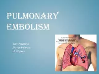
Pulmonary Embolism. Victor Politi, M.D.,FACP Medical Director SVCMC- School of Allied Health. Introduction. Thrombophlebitis - Inflammation of the vein with the presence of a blood clot. It can be difficult to know what came first — the clot or the inflammation ?. Introduction.

E N D
Pulmonary Embolism Victor Politi, M.D.,FACP Medical Director SVCMC- School of Allied Health
Introduction • Thrombophlebitis - Inflammation of the vein with the presence of a blood clot. • It can be difficult to know what came first — the clot or the inflammation ?
Introduction • A blood clot is a jelly-like mass of congealed blood. • Clotting is the normal way the body stops bleeding and begins healing following injury. • Once the clot has done its job, the body absorbs it. • Sometimes, however, blood clotting can prove harmful.
Introduction • Deep vein thrombosis (DVT) is a condition in which blood clots form in a vein deep within the body. The word thrombosis means forming a blood clot. The clot itself is called a thrombus
Patients who have undergone gynecologic surgery, those with major trauma, and those with indwelling venous catheters may have DVTs that start at any location. • For other patients, lower extremity venous thrombosis nearly always starts in the calf veins, which are involved in virtually 100% of all cases of symptomatic spontaneous lower extremity DVT. • Although DVT starts in the calf veins, it already has propagated above the knee in 87% of symptomatic patients before the diagnosis is made.
Introduction • DVT usually involves the formation of a large clot in the deep veins in the lower legs and thighs. • In rare instances, DVT can occur in the area around the armpit and collar bone (axillary-subclavian vein thrombosis), in the upper arm, abdomen, or pelvic region.
Introduction • Deep vein thrombosis most often occurs in: • Hospitalized patients following surgery. • Individuals confined to bed for prolonged periods. • Healthy individuals whose legs remain immobilized for long stretches of time, such as passengers on lengthy airline flights.
Introduction • Some people are more likely than others to develop thrombosis. Those at risk include: • The elderly • Diabetics • People with blood disorders • Women who take oral contraceptives (birth control pills) or other medications that contain the hormone estrogen • People with a history of thrombosis • People who have just undergone major surgeries or have just suffered a bone fracture
Introduction • Deep vein thrombosis is the second most common vascular problem in the United States. (The first is varicose veins.) • The condition is most commonly seen in people over age 60, but anyone can be affected
DVT is a dangerous condition because the clot may become dislodged from the vein and travel inside the vein to the lung, where it may get trapped and block a vessel in the lung. • This is called pulmonary embolism, which can be deadly.
Pulmonary embolism (PE) is an extremely common and highly lethal condition that is a leading cause of death in all age groups.
PE FACTS • It is the first or second most common cause of unexpected death in most age groups. • Nine out of 10 cases of pulmonary embolism are caused by blood clots that form in the legs and then travel to the lungs. • 3rd most common cause of death in the US. • More than 600,000 people in the United States have a pulmonary embolism each year, and more than 10 percent of them die from it. • Most who die do so within 30 to 60 minutes after symptoms start.
PE Facts • DVT of the calf is a significant source of PE and often causes serious morbidity or death. • In fact, 1/3 of the cases of massive PE have their only identified source in the veins of the calf.
Prompt diagnosis and treatment can dramatically reduce the mortality rate and morbidity of the disease. • Unfortunately, the diagnosis is missed far more often than it is made, because PE often causes only vague and nonspecific symptoms.
Introduction • Because PE is both extremely common and fairly difficult to diagnose, many patients are seen in the ED and later die from undiagnosed PE. • In fact, respiratory complaints are the most common complaints in patients who are seen alive in the ED and later die unexpectedly
Pathophysiology • Pulmonary thromboembolism is not a disease in and of itself. • It is an often fatal complication of underlying venous thrombosis.
Pathophysiology • Under normal conditions, microthrombi (tiny aggregates of red cells, platelets, and fibrin) are formed and lysed continually within the venous circulatory system. • This dynamic equilibrium ensures local hemostasis in response to injury without permitting uncontrolled propagation of clot.
Pathophysiology • Under pathological conditions, microthrombi may escape the normal fibrinolytic system to grow and propagate. • PE occurs when these propagating clots break loose and embolize to block pulmonary blood vessels.
Pathophysiology • Abnormal thrombus formation can be caused by either: • stasis of blood flow • hypercoagulability of the blood • or damage to the endothelial lining of the veins. • These 3 underlying causes are known as the Virchow triad. • All known clinical risk factors for DVT and PE have their basis in one or more of the triad.
Virchow’s triad • Thrombosis in the veins is triggered by venostasis, hypercoagulability, and vessel wall inflammation. Conditions that can cause venous stasis are: • varicose veins, obesity, surgery, prolonged bed rest, CHF and pregnancy.
Virchow’s triad • PE is the most common cause of maternal death. The risk of venous thrombo-embolism is 6× greater in pregnant women.
Virchow’s triad • Conditions that may cause hypercoagulation are: • malignant neoplasms, blood diseases that raise the platelet count, decreased fibrinolysis, inherited coagulopathies, dehydration, increase in the clotting factors that increase blood viscosity, and oral contraceptives or drugs that may enhance clotting.
Virchow’s triad • Conditions that may cause endothelial injury are: • IV injections, severe trauma and major orthopedic injury or surgery, contrast media for x-rays, and certain antibiotics.
Virchow’s triad • Conditions that may cause vascular wall injury are: • severe trauma with major orthopedic injury, major surgery, indwelling IV catheters, injections of irritating substances such as contrast media and certain antibiotics, IV drug abuse and prior deep vein thrombosis.
Virchow’s triad • Signs and symptoms of venous thromboembolic disease may include: • chronic edema • pain (claudication) • altered pigmentation • induration
Frequency • Surgical patients have long been recognized to be at special risk for DVT and PE, but the problem is not confined to surgical patients. • PE is the most common cause of maternal death. The risk of venous thrombo-embolism is 6× greater in pregnant women.
Mortality/Morbidity • Massive PE is one of the most common causes of unexpected death • second only to coronary artery disease as a cause of sudden unexpected natural death at any age • The diagnosis is unsuspected until autopsy in approximately 80% of cases.
Mortality/Morbidity • Although PE often is fatal, prompt diagnosis and treatment can reduce the mortality rate dramatically. • Approximately 10% of patients in whom acute PE is diagnosed die within the first 60 minutes. • Of the remainder, the condition eventually is diagnosed and treated in one third and remains undiagnosed in two thirds. • .
Mortality/Morbidity • Patients who survive an acute PE are at high risk for recurrent PE and for the development of pulmonary hypertension and chronic cor pulmonale right vent hypertrophy and failure • This occurs in up to 70% of patients and carries its own attendant mortality and morbidity
History • PE is so common and so lethal that the diagnosis should be sought actively in every patient who presents with any chest symptoms that cannot be proven to have another cause
History • Symptoms that should provoke a suspicion of PE must include: • chest pain, chest wall tenderness, back pain, shoulder pain, upper abdominal pain, syncope, hemoptysis, shortness of breath, painful respiration, new onset of wheezing, any new cardiac arrhythmia, or any other unexplained symptom referable to the thorax
History • The classic triad of signs and symptoms of PE (hemoptysis, dyspnea, chest pain) are neither sensitive nor specific. • They occur in fewer than 20% of patients in whom the diagnosis of PE is made, • Most patients with those symptoms are found to have some etiology other than PE to account for them.
History • Of patients who go on to die from massive PE: • 60% have dyspnea • 17% have chest pain • 3% have hemoptysis • Many patients with PE are initially completely asymptomatic, and most of those who do have symptoms have an atypical presentation
History • These patients usually lack any other classical signs, symptoms, or known risk factors for pulmonary thromboembolism. • Such patients often are dismissed inappropriately with an inadequate workup and a nonspecific diagnosis, such as musculoskeletal chest pain or pleurisy.
Physical • Massive PE causes hypotension due to acute cor pulmonale • Physical examination findings early in submassive PE may be completely normal. • Initially, abnormal physical findings are absent in most patients with PE.
Physical • After 24-72 hours, loss of pulmonary surfactant often causes atelectasis and alveolar infiltrates that are indistinguishable from pneumonia on clinical examination and by x-ray
Causes: Hypercoagulable states • Prolonged venous stasis or significant injury to the veins can provoke DVT and PE in any person, • increasing evidence suggests that spontaneous DVT and PE nearly always are related to some underlying hypercoagulable state. • Other identified "causes" most likely serve only as triggers for a system that is already out of balance.
Causes: Hypercoagulable states • Hypercoagulable states may be acquired or congenital. • An inborn resistance to activated protein C is the most common congenital risk factor for DVT that has been identified to date. • Most patients with this syndrome have a genetic mutation in factor V known as "factor V Leyden," although other mechanisms also can produce a resistance to activated protein C
Causes: Hypercoagulable states • Primary or acquired deficiencies in protein C, protein S, or antithrombin III are also common underlying causes of DVT and PE
Risk Markers • The most important clinically identifiable risk markers for DVT and PE are: • prior history of DVT or PE • recent surgery or pregnancy • prolonged immobilization • underlying malignancy / Trousseau’s syndrome= cancer +recurrent superficial and Deep Venous Thrombosis
Other recognized markers of risk for venous thromboembolic disease • AIDS (lupus anticoagulant) • Antithrombin III deficiency • Behçet disease • Blood type A • Burns • Catheters (indwelling venous infusion catheters) • Chemotherapy • Congestive heart failure (CHF) • Drug abuse (intravenous [IV] drugs) • Drug-induced lupus anticoagulant • DVT in the past • Estrogen replacements (high dose only)
Other recognized markers of risk for venous thromboembolic disease • Fibrinogen abnormality • Fractures • Hemolytic anemias • Heparin-associated thrombocytopenia • Homocystinuria • Hyperlipidemias • Immobilization • Malignancy • Myocardial infarction • Obesity • Old age
Other recognized markers of risk for venous thromboembolic disease • Oral contraceptives • PE in the past • Phenothiazine • Plasminogen abnormality • Plasminogen activator abnormality • Polycythemia • Postoperative • Postpartum period • Pregnancy • Protein C deficiency • Protein S deficiency • Resistance to activated protein C • Systemic lupus erythematosus
Other recognized markers of risk for venous thromboembolic disease • Thrombocytosis • Trauma • Ulcerative colitis • Varicose veins • Venography • Venous pacemakers • Venous stasis • Warfarin (first few days of therapy)
Other Problems to be Considered • Whether the presentation of the patient with pulmonary thromboembolism is typical or atypical, the list of differential diagnoses remains extensive and the true diagnosis must be sought actively
Diagnostic Work-Up • Clinical variables alone lack sufficient power to permit a treatment decision • patients in whom PE is suspected must undergo diagnostic tests until the diagnosis is proven or ruled out, or until some alternative diagnosis is proven. • no known blood or serum test can move a patient with a high clinical likelihood of pulmonary thromboembolism into a low likelihood category or vice versa
Lab Studies • The PO2 on arterial blood gases analysis (ABG) has a zero or even negative predictive value in a typical population of patients in whom PE is suspected clinically • Clotting study results are normal in most patients with pulmonary thromboembolism • The white blood cell (WBC) count may be normal or elevated. • A WBC count as high as 20,000 is not uncommon in patients with PE
Lab Studies • At the present time, D-dimer is not sensitive or specific enough to change the course of diagnostic evaluation or treatment for patients with suspected PE • A-a gradient PAO2-PaO2 • PAO2=150-(1.25xPCO2) use PaCo2 from the ABG for PCO2 • Assumes room air FiO2=21%@sealevel • Normal 10-15
Radiologic Studies - CXR • initial chest x-ray (CXR) findings of a patient with PE are virtually always normal • Rarely may show Westermark sign – • dilatation of the pulmonary vessels proximal to an embolism along with collapse of distal vessels, sometimes with a sharp cutoff. • Over time, an initially normal CXR often begins to show atelectasis • which may progress to cause a small pleural effusion and an elevated hemidiaphragm.