Download
1 / 38
400 likes | 763 Vues
Unit 3 Autoimmunity Part 2 Systemic Lupus Erythematosus Part 3 Rheumatoid Arthritis. Terry Kotrla, MS, MT(ASCP)BB. Expectation. Students are expected to know: Signs and symptoms, especially part of body affected Age and sex if appropriate Tests to diagnose Treatment.

E N D
Unit 3 AutoimmunityPart 2 Systemic Lupus ErythematosusPart 3 Rheumatoid Arthritis Terry Kotrla, MS, MT(ASCP)BB
Expectation • Students are expected to know: • Signs and symptoms, especially part of body affected • Age and sex if appropriate • Tests to diagnose • Treatment
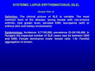
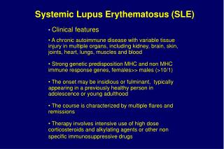
Systemic Lupus Erythematosus • Chronic, systemic inflammatory disease caused by immune complex formation. • The word "systemic" means the disease can affect many parts of the body. • Pathophysiology associated with clinical features secondary to immune complexes depositing in tissues resulting in inflammation. • Parts of the body affected include: the joints, skin, kidneys, heart, lungs, blood vessels, and brain.
Systemic Lupus Erythematosus • Peak age of onset is 20 to 40 years of age. • Found more frequently in women. • Has both genetic and environmental factors. • Often difficult to diagnose. • “Great imitator” as it mimics or is mistaken for other illnesses. • Can be fatal but survival rates increasing.
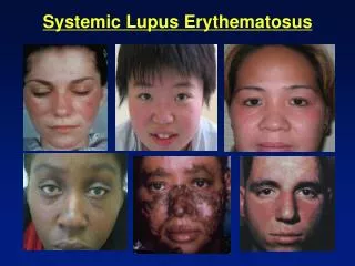
SLE Clinical Signs • Extremely diverse and nonspecific. • Joint involvement most frequent signs are polyarthralgia and arthritis which occur in 90% of patients. • Skin manifestations next most common. • Erythematosus rash may appear. • Most classic is butterfly rash.
SLE Butterfly Rash • The source of the name "lupus" is unclear. All explanations originate with the characteristic butterfly-shaped malar rash that the disease classically exhibits across the nose and cheeks. • Various accounts, some doctors thought the rash resembled a wolf pattern. In other accounts doctors thought that the rash, which was often more severe in earlier centuries, created lesions that resembled wolf bites or scratches. • Stranger still, is the account that the term "Lupus" didn't come from latin at all, but from the term for a French style of mask which women reportedly wore to conceal the rash on their faces
SLE Clinical Signs • Renal involvement very common. • Caused by deposition of immune complexes in kidney tissue. • Leads to renal failure, most common cause of death. • Other systemic effects: • Cardiac • Central nervous system • Liver • Hematologic abnormalities
Immunologic Findings • Lupus Erythematosus (LE) cell, neutrophil which has engulfed the antibody-coated nucleus of another cell. • First classic test to aid in diagnosis. • Not utilized anymore, may still see in older references. • Over activity of B cells main immunologic characteristic. • Antinuclear antibodies produced. • More than 28 antibodies associated with LE have been identified. • Level of antibody production correlates with severity of symptoms. • Estrogen enhance B cell activation.
LE Cell • "LE cell" test which has value only in demonstrating how the concept of autoantibodies work. • Pink blobs are denatured nuclei. • Two in this slid, one being phagocytosed in the center by a PMN. • This test is not nearly as sensitive as the ANA which has supplanted the LE cell test. Therefore, NEVER order an LE cell test. [Image contributed by Elizabeth Hammond, MD, University of Utah]
Immunologic Findings • Decrease in absolute number of T cells • Accumulation of immune complexes with activation of complement lead to kidney damage. • Drug induced lupus may occur, discontinue drug, symptoms usually disappear.
Laboratory Diagnosis • Screening test for anti-nuclear antibodies (ANA) first test done. • Antibodies directed against nuclear material of cells. • Flourescent anti-nuclear antibody (FANA) most widely used, extremely sensitive, low diagnostic specificity. • Animal or human cells fixed to slide. • Add patient serum and incubate. • Wash to remove unreacted antibody. • Add anti-human globulin labeled with fluorescent tag or enzyme.
Antinuclear Antibody Test • Antinuclear antibodies (ANA) are autoantibodies against various cell nucleus antigens and are found in patients with autoimmune diseases such as SLE. • Some of ANA are considered to be useful for diagnosis of autoimmune diseases. • This picture illustrates the most common antigens used in the ANA • At the MLT level you will not be required to memorize.
ANA • Patients antinuclear antibody titer of 1:40 and characteristic multiorgan system involvement can be diagnosed with SLE without additional testing • Patients with antibody titer of 1:40 who fail to meet full clinical criteria should undergo additional testing including: • Tests for antibody to doublestranded DNA antigen • Antibody to Sm nuclear antigen. • Antinuclear antibody titer of less than 1:40 usually rules out systemic lupus erythematosus but patients with persistent, characteristic multisystem involvement may be evaluated for possible antinuclear antibody–negative disease.
ANA • Patterns of reactivity: • Homogenous-entire nucleus stained • Peripheral-rim of nucleus stained • Speckled-spots of stain throughout nucleus • Nucleolar-nucleolus only stained • False positives and negatives occur. • If positive, perform profile testing.
ANA • For the next exam you must be able to: • Name the 4 primary reactions • Describe the 4 primary reactions seen • Identify the type of reaction in a photo
Homogeneous Pattern • Smooth, even staining of the nucleus with or without apparent masking of the nucleoli
Nucleolar • 23 or 46 (or some multiple of 46) bright speckles or ovoid granules spread over the nucleus of interphase cells
Peripheral • Fluorescence is most intense at the periphery of the nucleus with a large ring starting from the internal nuclear membrane and the rest of the nucleus showing weaker yet smooth staining.
Speckled • Large speckles covering the whole nucleoplasm, interconnected by a fine fluorescent network.
Anti-nuclear antibodies detected by FANA • Double-stranded DNA (ds-DNA) antibodies are most specific for SLE, correlate well with disease activity. • Antihistone antibody second major antibody found in SLE. • Deoxyribonucleoprotein (DNP) antibody, responsible for LE cell phenomena and available as a latex agglutination test. • Anti-Sm antibody, specific for LE. • SS-A/Ro and SS-B/La antibodies, most common in patients with cutaneous manifestations. • Anti-nRNP detected in patients with SLE as well as mixed connective tissue disease. • Presence of antibodies not diagnostic, may be present due to other diseases.
Anti-Nuclear Antibody by Immunodiffusion • Used to determine specificity. • Ouchterlony double diffusion most frequently used to identify antibodies to: Sm, nRNP, SS-A/Ro, SS-B/La and others. • Test is not as sensitive but very specific.
Extractable Nuclear Antigen • Antibody to cytoplasmic and nuclear components. • Over 100 different antigens described. • It is associated with mixed connective disease and SLE with particular features (arthritis, myositis, Raynaud's phenomenon - also association with HLA-DR4 and HLA-DQw8).
Antiphospholipid Antibodies • Antiphospholipid antibodies may be present and are of two types. • Anticardiolipin. • Lupus anticoagulant, if present, may cause spontaneous abortion and increase • Risk of clotting, platelet function may be affected.
Treatment • Aspirin and anti-inflammatories for fever and arthritis. • Skin manifestations-anti-malarials or topical steroids. • Systemic corticosteroids for acute fulminant lupus, lupus nephritis or central nervous system complications. • Five year survival rate is 80 to 90%.
Rheumatoid Arthritis • Chronic systemic inflammatory disease primarily affecting the joints, but can affect heart, lung and blood vessels. • Women three more times as likely as men to have it. • Typically strikes at ages between 20 and 40, but can occur at any age. • The three major symptoms of arthritis are joint pain, inflammation, and stiffness. • Progress of disease varies.
Arthritis • Group of conditions involving damage to the joints of the body. • Over 100 different forms of arthritis. • Will discuss the autoimmune type, rheumatoid arthritis.
Clinical Signs • Diagnosis based on criteria established by American College of Rheumatologists, must have at least 4 of the following: • Morning stiffness lasting 1 hour. • Swelling of soft tissue around 3 or more joints. • Swelling of hand/wrist joints. • Symmetric arthritis. • Subcutaneous nodules • Positive test for rheumatoid factor. • Xray evidence of joint erosion.
Clinical Signs • Symptoms initially non-specific: malaise, fever, weight loss, and transient joint pain. • Morning stiffness and joint pain improve during the day. • Symmetric joint pain: knees, hips, elbows, shoulders. • Joint pain leads to muscle spasm, limits range of motion, results in deformity. • Approximately 25% of patients have nodules over bones (necrotic areas), nodules can also be found in organs. • Certain bacteria may trigger RA due to certain proteins that possess antigens similar to those antigens found in joint, ie, molecular mimicry
Immunologic Findings • Rheumatoid Factor (RF) is an IgM antibody directed against the Fc portion of the IgG molecule, it is an anti-antibody. • Not specific for RA, found in other diseases. • Immune complexes form and activate complement and the inflammatory response. • Enzymatic destruction of cartilage is followed by abnormal growth of synovial cells, results in the formation of a pannus layer.
Diagnosis • Diagnosis is based on: • Clinical findings. • Radiographic findings • Laboratory testing.
Laboratory Testing • Rheumatoid Factor • IgM autoantibody directed against the Fc portion of the antibody molecule. • Detected by testing patient serum with red blood cells or latex particles coated with IgG, agglutination is a positive result. • Nephelometry and ELISA techniques are available to quantitate the RF. • Erythrocyte Sedimentation Rate (ESR) used to monitor inflammation. • C-Reactive protein (CRP) is utilized to monitor inflammation
Treatment • Goal to achieve lowest level of disease, remission if possible, minimization of joint damage. • Rest and non-steroidal anti-inflammatory drugs control swelling and pain. • Substantial functional loss seen in 50% of patients within 5 years. • Slow acting anti-rheumatic drugs are coming into use but have side affects. • Joint replacement.
The End • Write a question about anything you did not understand in this unit. • You may choose to ask a question about any of the presentations required for viewing the next class period. • WRITE YOUR ANSWERS TO THE 6 QUESTIONS presented in this presentation on a sheet of paper and submit when you walk in the door.