Download
1 / 24
270 likes | 493 Vues
Cardiac Cycle Setting the Tempo. Cardiac Cycle – Overview. Events of each heartbeat Highly coordinated so that both atria contract together and then both ventricles contract together Systole contraction of heart muscle Diastole relaxation of heart muscle. Overview cont’d.

E N D
Cardiac Cycle – Overview • Events of each heartbeat • Highly coordinated so that both atria contract together and then both ventricles contract together • Systole • contraction of heart muscle • Diastole • relaxation of heart muscle
Overview cont’d • Time for each cycle is influenced by autonomic nerves. • Two regulatory nervous systems • Sympathetic nervous system • Forms a division of the autonomic nervous system • Prepares the body for stress increases heart rate. • Parasympathetic nervous systems • Returns the body to normal resting state following stress
Cardiac Cycle cont’d • Cardiac muscles are able to contract without being stimulated by external nerves. • Called myogenic muscle • allows your heart to beat without a continual reminder • Also able to recover quickly, the only rest they get is small time between beats. • Normal heart rate at rest is about 60-80 beats per minute
Stages of the Cardiac Cycle • Fig 12.5
Caused by the closing of the heart valves Lubb – AV valves close Dubb – Semi lunar valves close Heart murmurs are caused by an incomplete seal on a valve Blood leaks past the closed valve, or flows backwards in the heart. causes a whoosing, or gurgling sound Heart Beat - Sounds
Heart Beat - Intrinsic Control • Heart has its own intrinsic conduction system • Autorhymicity-unlike skeletal muscle, cardiac muscle can contract without neural stimulation • The autonomic nervous system does has inputs to the heart and normally regulates rate • Nodal tissue-2 areas in the heart • Has both muscular and nervous characteristics • Can generate action potentials to cause contraction • SA node and AV node
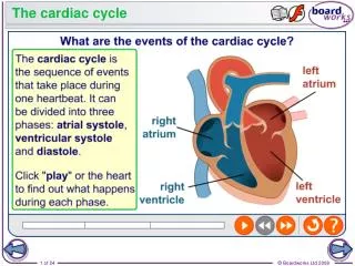
Sinoatrial Node (SA) A bundle of specialized nerves and muscles located where the vena cava enter the right atrium. (#1) Sends a signal over the two atria Atrioventricular node (AV). Located the lower part of the right atrium close to the tricuspid valve (#2) picks up electrical impulses Heart Beat – The Pacemaker
Heart Beat – The Pacemaker • AV Node sends nerve impulses via the Purkinje Fibres (#4) • two large nerve fibres • run through the septum, • Each nerve impulse triggers cardiac contraction • Atria contract first, followed by ventricles.
Heart Beat • Tachycardia • Fast heart beat • heart rate exceeds 100 beats per min • can result from exercise or from the consumption of such drugs as caffeine or nicotine. • Bradycardia • Slow heart beat • Heart beat lower than 60 bpm • Can result from degeneration of the muscle (age), disease
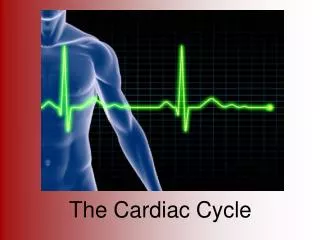
Mapping the Heart Beat Electrocardiogram (ECG or EKG) test that measures the electrical activity of the heart. Traces how long the electrical wave takes to pass through your heart Printed on paper covered with a grid of squares each represents 0.04 seconds. 25 squares = 1 second ECG’s printouts are usually is 6 seconds; a "six second strip." Changes in electrical current reveal normal or abnormal events of the cardiac cycle. Determines if activity is normal, fast or irregular. heart is enlarged or overworked.
Interpreting ECG • P wave • first little hump – atria receives signal and contracts • QRS complex • Ventricles receive signal and contract • May just be an RS… this is still normal • R wave is the first wave ABOVE the midline • T wave • Recovery of the heart
Abnormal Rhythym Normal Rhythm Bradycardia Tachycardia V-Tach (NO QRS)
Blood Pressure • A measure of the pressure or force of blood against the walls of your arteries • Systolic • the pressure when your heart contracts and pushes blood out • Highest pressure • Diastolic • the lowest pressure when the heart relaxes between beats • Normal blood pressure is below 120/80 mm Hg. • High blood pressure is consistently more than 140/90 mm Hg
Our Heart the Pump Diastole Systole
Sphygmomanometer • Measures blood pressure indirectly • Measures the pressure exerted by blood in the brachial artery • Blood flowing through the brachial artery makes no noise • A stethoscope is placed below the cuff to listen for the blood • The cuff is placed around your arm and brachial artery. • Air is pumped into the cuff until circulation is restricted.
Sphygmomanometer • Pressure is slowly lowered until the blood is able to flow past, this is the systolic pressure • Doctors will hear this blood rushing through as a beat • Pressure continues to be lowered until the noise disappears • This is the diastolic pressure • Blood no longer needs to force the artery open to pass through.
Factors affecting Blood Pressure • Cardiac output • The volume of blood pumped from the heart each minute • Increased output will increase blood pressure. • Raising your heart rate increases output!
Factors affecting Blood Pressure • Arteriolar resistance • Diameter of the arterioles is regulated by muscles in their walls • Arteriolar Constriction reduces blood flow, causing higher blood pressure. • Arteriolar dilation, opens vessels increases blood flow and decreases blood pressure
High Blood Pressure Factors • High Blood Volume • High Salt levels cause excess water in our blood increasing our blood volume • Increased Cardiac Output • Tachycardia • Overactive Sympathetic Nervous System • Arteriolar Constrictors • Caffeine and Nicotine • Cold • Blockages in the arteries • Caused by artheriosclerosis
Low Blood Pressure Factors • Low Blood Volume • Dehydration • Starvation (anorexia) • Bleeding • Decreased Cardiac Output • Bradycardia • Insufficient stroke volume • Valve problems • Arteriolar Dilation • Heat • Brain injury • Arteriolar walls stop contracting