Download
1 / 32
340 likes | 719 Vues
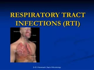
Infections of the Respiratory Tract. Dr. Raid Jastania. Infections of the Respiratory Tract. Upper Respiratory Tract Lower Respiratory Tract Bacterial, Viral, Fungal, T.B, Parasitic Most URT infections are viral Most LRT infections are bacterial. Upper Respiratory Tract Infections.

E N D
Infections of the Respiratory Tract Dr. Raid Jastania
Infections of the Respiratory Tract • Upper Respiratory Tract • Lower Respiratory Tract • Bacterial, Viral, Fungal, T.B, Parasitic • Most URT infections are viral • Most LRT infections are bacterial
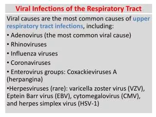
Upper Respiratory Tract Infections • Common cold (Acute coryza) • Viral infection of URT • Organisms: • Rhinoviruses: Coronaviruses, Enteroviruses, Adenoviruses, Respiratory syncytial virus) • Influenza A and B • Croup (Parainfluenza 1,2,3)
Upper Respiratory Tract Infections • Tonsillitis (mostly bacterial) • Otitis media (mostly bacterial) • Epiglottitis • Laryngitis • Laryngotrachiobronchitis • Bronchitis • Bronchiolitis • Pneumonia
Pneumonia • Pneumonia is inflammation of the lung (lower respiratory tract) caused mainly by infection. • Pneumonia can be caused by Bacterial infection and less commonly by other organisms eg. Viruses, Fungi • The term Pneumonia is sometimes used to indicated inflammation of lungs due to other causes eg. Including interstitial lung disease (interstitial pneumonia)
Types of Pneumonia • Different ways of classification • Problematic, confusing • Classification is Based on • etiology, • anatomic site involved, • clinical presentation, • pathological type of inflammation
Types of Pneumonia • One of the classification divides pneumonia into: • Primary (community-acquired) • Secondary • Others
Types of Pneumonia • One of the classification divides pneumonia into: • Primary (community-acquired) • Typical pneumonia • Lobar pneumonia • Bronchopneumonia • Atypical pneumonia • Secondary • Aspiration pneumonia • Nosocomial (hospital-acquired) pneumonia • Pneumonia in immunosuppression • Others: • Chronic pneumonia • Necrotizing pneumonia/Supporative pneumonia/Lung Abscess
Risk of Pneumonia • Underlying disease • COPD • Heart failure • Diabetes • Immunodeficiency • Absent splenic function (sickle cell disease)
Clinical Presentation • Fever, rigor, malaise, weakness, vomiting, loss of appetite, headache • Cough with sputum • Dyspnea • Chest pain, pleuritic pain • Sick, ill , distressed • High respiratory rate >30 / mint • In lobar pneumonia: localized area of dullness on percussion, increased tactile fremitus, bronchial breath sounds, and crepitation, pleural rub
Morphology • Common in lower lobes and right middle lobe • In Lobar pneumonia: there is a localized area of inflammation • Stages: • Congestion • Vascular congestion, edema, few neutrophils • Red hepatization • Fibrin, RBC, neutrophils in alveolar spaces • Gray hepatization • Fibrin, RBC lysis • Resolution
Bronchopneumonia • Inflammation of the bronchi and bronchioles with collapse of the distal airspaces • Multiple, patchy bilateral small infiltrates • Affect lower lobes usually
Outcome and complications • Resolution • Fibrosis • Abscess • Empyema • Dissemination of infection • Meningitis, arthritis, endocarditis
Investigations • CBC • Arterial blood gases • Radiological exam: chest x-ray • Sputum exam and culture • Nose and throat swabs • Blood culture • Serological tests
Pneumonia: Features of different organisms (community-acquired pneumonia) • Strep. Pneumoniae • commonest • Staph. Aureus • Common following viral infection • Risk of complications: abscess • Common in IV drug abusers • Legionella • Legionnaire’s disease, epidimics • Grow in water reservoir, humidifiers • People with heat disease, renal disease, immunosuppressed • Presentation with GIT symptoms, mental confusion • Hemophilus influenzae • Common in COPD, chronic bronchitis, bronchiectasis, cystic fibrosis • Klebsiella • Chronic alcoholics and malnourished persons
Atypical Pneumonia • Viruses, Mycoplasma, Chlamydia • Fever and malaise precede the respiratory symptoms by few days • Severe headache, malaise, anorexia • No localized sings on chest exam, No consolidation on chest x-ray • Spleen may be enlarged • WBC normal, cultures negative • No improvement with Penicillin
Atypical Pneumonia (community-acquired) • Mycoplasma • Sporadic or epidemics • Viruses • Influenza, Parainfluenza, Adenovirus, respiratory syncytial virus, measles, chicken pox • Chlamydia
Atypical pneumonia • Morphology: • Patchy or involve whole lobe • Inflammation is confined to the alveolar walls • Widening of alveolar walls by edema, mononuclear cell infiltration (lymphocytes, plasma cells, macrophages)
Secondary pneumonia • Aspiration pneumonia • Nosocomial (hospital-acquired) pneumonia • Pneumonia in immunosuppression
Secondary Pneumonia • Pre-existing disease of lung or factors increasing the risk of infection • Low virulence organisms: Hemophilus infleunzae, viruses, fungi • Anaerobic bacteria • Gram negative bacteria • Staph aureus • All the others in commuity-acquired
Aspiration Pneumonia • Aspiration of gastric contents • During surgery, anesthesia, surgery of tonsils, dental work • Infection following Aspiration of vomitus in coma, anesthesia, or sleep • Ineffective coughing (post operative) • Can result in severe hemorrhage in lungs • Chemical injury + infection (Anaerobic) • Destruction of lung parenchyma with cavitations
Nosocomial Pneumonia • Patients admitted to hospital • Organisms • Same as community acquired and • Gram-negative (Klebsiella, E.coli, Pseudomonas) • Staph. Aureus
Pneumonia in Immunosuppression • Congenital or acquired • AIDS, Immunosuppression • Humoral and Cellular immunity • Infection by • Pneumocystis carinii • Gram negative bacteria • The common bacteria • Opportunistic pathogens: CMV, Herpes, Aspergillus, TB, mycobacteria
Lung Abscess • Suppurative pneumonia • Necrotizing pneumonia • Cavity • Localized suppurative necrosis
Lung Abscess • Mechanisms: • Aspiration of infective material: teeth, tonils, coma, alcoholics • Aspiration of gastric conetnets • Complication of necrotizing pneumonia • Bronchial obstruction • Septic emboli • Hematogenous spread
Lung Abscess • Morphology • Cavity 1-2mm to 5-6 cm • Filled with pus, cellular debris • Surrounded by fibrosis and chronic inflammation • Aspiration tend to involve the right lung • May rupture in airways resulting in Air-fluid levels • May rupture in pleura resulting in pneumothorax and empyema