Download
1 / 13
130 likes | 203 Vues
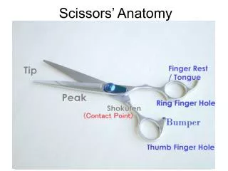
Cut away fat using scissors. Anterior External Eye Structure. Posterior External Eye Structures.

E N D
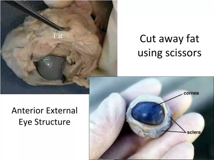
Cut away fat using scissors Anterior External Eye Structure
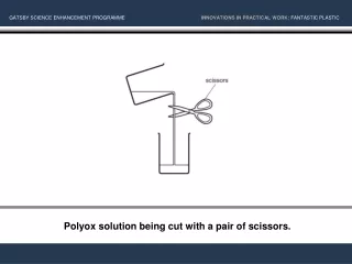
Select a place to make an incision of the sclera midway between the cornea and optic nerve. Use the point of surgical scissors to make a small cut through the sclera. Fluid should ooze out of the eyeball when you have cut deeply enough.
Arrange the two hemispheres of the eye as you see in the photograph. • Observe the semi-fluid vitreous humor that fills the central cavity of the eye. It is transparent in the living eye but might be cloudy in the preserved specimen
The retina lines the posterior cavity of the eye and extends forward to the ciliary body. Use your probe to lift and pull the retina back from the underlying choroid layer. • Notice that the retina is only firmly attached to the choroid at one place. This region is the optic disc or blind spot.
Internal Eye Structure – Posterior Section tapetum lucidum: the iridescent reflecting tissue layer of the choroid of some species of animals that gives their eyes the property of shining in the dark. It is characteristic of nocturnal animals and allows incident light two opportunities to stimulate the retinal receptors. Called also choroidal tapetum.
Remove the lens and place against newspaper to see that it is a magnifier!
When the lens is removed, an opening, allowing light to enter the eye is seen. This opening, the pupil is located in the center of the iris. Note the oblong shape of the sheep pupil, in humans the pupil is circular. • The back side of the iris can be seen just above the pointer in the photograph.