Download
1 / 41
410 likes | 567 Vues
The effects of ultraviolet B treatment on the expression of adhesion molecules by circulating T lymphocytes in psoriasis. Presented By: Anika Ramos. OUTLINE. 1. Introduction 2. What is psoriasis 3. What causes it? 4. T-cells… 5. Genetically Predisposed? 6. Cytokines

E N D
The effects of ultraviolet B treatment on the expression of adhesion molecules by circulating T lymphocytes in psoriasis Presented By: Anika Ramos
OUTLINE • 1. Introduction • 2. What is psoriasis • 3. What causes it? • 4. T-cells… • 5. Genetically Predisposed? • 6. Cytokines • 7. Article Presentation
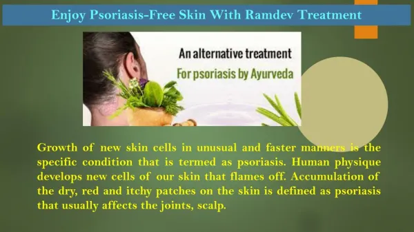
What is Psoriasis? • Psoriasis is a chronic (long-lasting) inflammatory dermatitis • It affects approximately 2% of the population(5.5 million people) • It is characterized by red, scaly skin and patches that are usually found on the scalp, elbows, and knees
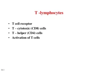
What causes Psoriasis? • Recent research indicates that psoriasis is a disorder of the immune system • The immune system includes a type of white blood cell, called a T cell. • T cells usually protect the body against infection and disease • However, in psoriasis an abnormal immune system causes negative activity by T cells in the skin
Function of Normal T Cells • There are two types of white blood cells 1. T- cells 2. B- cells • When T –cells identify a foreign material or organism, they attack it • When B-cells identify a foreign material, they secrete special chemicals called antibodies. • These antibodies stick on the foreign material and destroy it
What Happens in Psoriasis? • Psoriasis causes abnormal immune system activity of T-cells in the skin • These T-cells cause the skin to become inflamed and reproduce excessively • It is therefore called a T-cell mediated disease
What type of T-cells are involved? • The CD4+ T cells predominate in the dermis • The CD8+ T cells localize to the epidermis
Is Psoriasis a Disease With Genetic Predisposition? • Psoriasis is a heterogeneous disease in its clinical expression • Both genetic and environmental factors are thought to contribute to the pathogenesis • There is a higher than average incidence of psoriasis in relatives of people with psoriasis, indicating “familial tendency”
The Role of Cytokines in the Pathogenesis of Psoriasis • T-cells as well as cytokines are of major importance in the pathophysiology of psoriasis • Cytokines are a unique family of growth factors. • Secreted primarily from leukocytes, • cytokines stimulate cellular immune responses, as well as the activation of phagocytic cells.
Cytokines… • Basically, the over expression of these proinflammatory cytokines is considered to be responsible for initiation, maintenance, and recurrence of psoriatic skin lesions • What happens is that in contrast to the overexpression of proinflammatory cytokines, there is a relatively low level of expression of the antiinflammatory cytokines • What results is an insufficient counterregulatory capacity in psoriasis
The Effects of Ultraviolet B Treatment on the Expression of Adhesion Molecules by Circulating T lymphocytes in Psoriasis • Presented by Anika Ramos
Background… • T lymphocytes are believed to play a role in the pathogenesis of psoriasis • More than 80% of T lymphocytes that infiltrate psoriatic lesions express the surface glcoprotein called: • Cutaneous Lymphocyte Associated Antigen (CLA) CLA is a 200 kDa cell-surface glycoprotein • This compares with expression in the blood which is less than 20%
Purpose of experiment • The purpose of this study was to compare the effects of UVB treatment of psoriasis on the expression of CLA and several other surface markers expressed by circulating T lymphocytes.
A reminder… • T- lymphocytes are thought to have an important role in the pathogenesis of psoriasis, and agents that act selectively on T cells have a potent antipsoriatic effect
“Radiation” • Exposure to ultraviolet B is a common and effective treatment for psoriasis. • UVB radiation suppresses their proliferative responses to allogenic cells and mitogens • High UVB doses act both on T cells and antigen presenting cells • Lower doses selectively affect T cells, including suppression of their cytokine production and expression
Adhesion molecules… • Several adhesion molecules are involved in the migration of T cells from the blood into tissues • T cells that infiltrate the skin express a unique skin homing determinant called: • Cutaneous Lymphocyte Associated antigen (CLA): • It is a carbohydrate epitope interacting with endothelial E-selectin that facilitates the targeting of T cells to inflamed skin
E-Selectin… • The expression of E-selectin is low in normal skin • In psoriatic skin the expression of E-selectin increases dramatically • Therefore, causing a large attraction for T cells in the skin
So what’s important for the spread of psoriasis? • A large amount of localized T cells • Expressing the CLA antigen • The very late antigen (VLA)-4 • And its cell adhesion molecule
Materials and Methods… • Seven patients with active chronic plaque psoriasis were included in the study: • 4-men • 3-women • Age (17-65) • The patients were not on ant other treatment except for moisterizing ointments
Treatment… • They were treated daily with narrow-band (312 nm) UVB • Initial dose: 0.1 mJ cm • Gradually increased during the 4 weeks to • Max dose: 1.7 mJ cm • The patients were all evaluated for a PASI score
What is PASI? • PASI stands for: • Psoriasis Area and Severity Index score • Doctors use this type of scoring method to indicate the severity of the psoriasis in any given area of the body
Materials and Methods continued… • Blood was drawn simultaneously from all seven patients at the beginning of treatment and weekly thereafter. • This blood was to be tested for several factors
Antigens… • Antigens are macromolecules that elicit an immune response in the body. The most common antigens are proteins and polysaccharides. • Antigens were added to the blood samples • they are associated with the T cell antigen receptor. • Required for cell surface expression of and signal transduction by TCR. • Specific antigen added was: • Streptococcal antigen
Monoclonal Antibodies… • Humans (and mice) have the ability to make antibodies able to • recognize (by binding to) virtually any antigenic determinant (epitope) • to discriminate between even similar epitopes. • Antibodies used: • Anti-CD4, Anti-CD8, Anti CD4/PE, Anti CD8/PE, and a couple more
Isolation of Mononuclear blood lymphocytes • Peripheral blood mononuclear cells (PBMC) were isolated from heparinized blood by a density gradient • The cells were washed and resuspended
Immunofluorescence staining and flow symmetry…. • The (PMBC) blood cells were double-stained with solutions containing the monoclonal antibodies • The stained cells were immediately submitted to flow symmetry analysis using a special machine called a FACScan, used specifically for this purpose • Analysis of unstimulated and stimulated cells was performed usin a light scatter that included both small and large T lymphocytes. • Mean fluorescence intensity was assessed by flow symmetry using the soft ware
Stimulation of lymphocytes • After the correct T-cells were recovered, they were cultured together with 1000 ng mL of the streptococcal superantigens • Cultured lymphocytes were pulsed with tritiated thymidine • Proliferation was determined by thymidine incorporation as measured by spectroscopy • They were also stained for surface marker expression
Statistical analysis… • Significance was determined by the paired t-test • P< 0.05 was considered significant
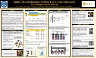
Results… • The mean PASI score had decreased from 13.4 to 3.5 (P= 0.006)\ • This correlated closely with a significant decrease in the intensity of CLA and VLA-4 expression by the T cells( P=0.016 and 0.015) • A corresponding but less marked decrease in the frequency of CLA T cells was observed • The frequency of VLA-4 T cells did not decrease during the treatment
Figure 1 • The mean PASI score decreases from 13.4 to 3.5 after 2 weeks of treatment • This correlated closely with a progressive and linear decrease in the intensity of CLA expression by the T cells
Figure 2 • As in the previous explanation, the expression of VLA-4 by T cells also decreased in correlation with the PASI score
Discussion • In this study they demonstrated a marked decrease in the expression by circulating T cells of the skin molecules CLA and VLA-4 during the first 2 weeks of UVB treatment • No decrease was observed in the expression of ICAM – 1 activation markers • Also lymphocyte responses to streptococcal antigens and superantigens remained unchanged
Conclusion • Treatment with narrow band UVB has been reported to deplete dermal T cells in psoriasis and to induce T-cell apoptosis in vivo • In view of this, the reduced expression of CLA and VLA-4 that was observed may be due to an elimination of T cells within the UVB exposed skin • It is an interesting possibility that sustained remission that is obtained after UVB treatment is due to apoptosis of T cell clones that are specific for autoepitopes in the skin