Download
1 / 5
50 likes | 59 Vues
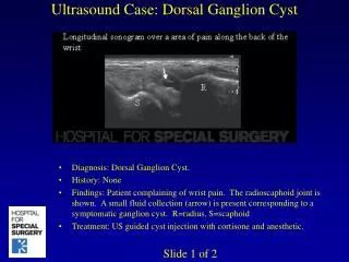
Solitary simple renal cyst is a cystic disease of kidney, incidentally found during routine abdominal imaging, dissection and autopsy without clinical significance. The prevalence of solitary renal cyst is increasing progressively with age, especially in the elderly population. These are asymptomatic but clinical features may result from rupture, haemorrhage or due to infection. Approximately 6 of solitary cysts are complicated by haemorrhage. Early diagnosis and careful follow up prevents complications. Cystic diseases of kidney are common, since many of these have effect on renal function, careful documentation is necessary. We describe cases of solitary renal cysts observed in a 70year old female and 55year male cadavers during routine dissection in the department of Anatomy. Vislavath Srikanth | Anju Thomas | Jyoti Umarji "Solitary Simple Renal Cyst: A Case Report" Published in International Journal of Trend in Scientific Research and Development (ijtsrd), ISSN: 2456-6470, Volume-2 | Issue-5 , August 2018, URL: https://www.ijtsrd.com/papers/ijtsrd17141.pdf Paper URL: http://www.ijtsrd.com/medicine/anatomy/17141/solitary-simple-renal-cyst-a-case-report/vislavath-srikanth<br>

E N D
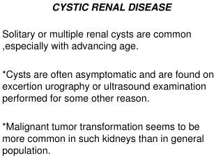
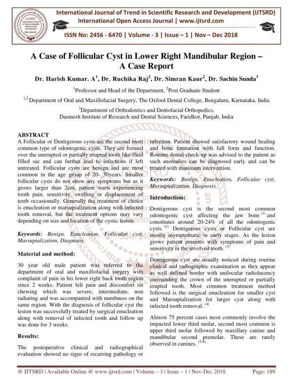
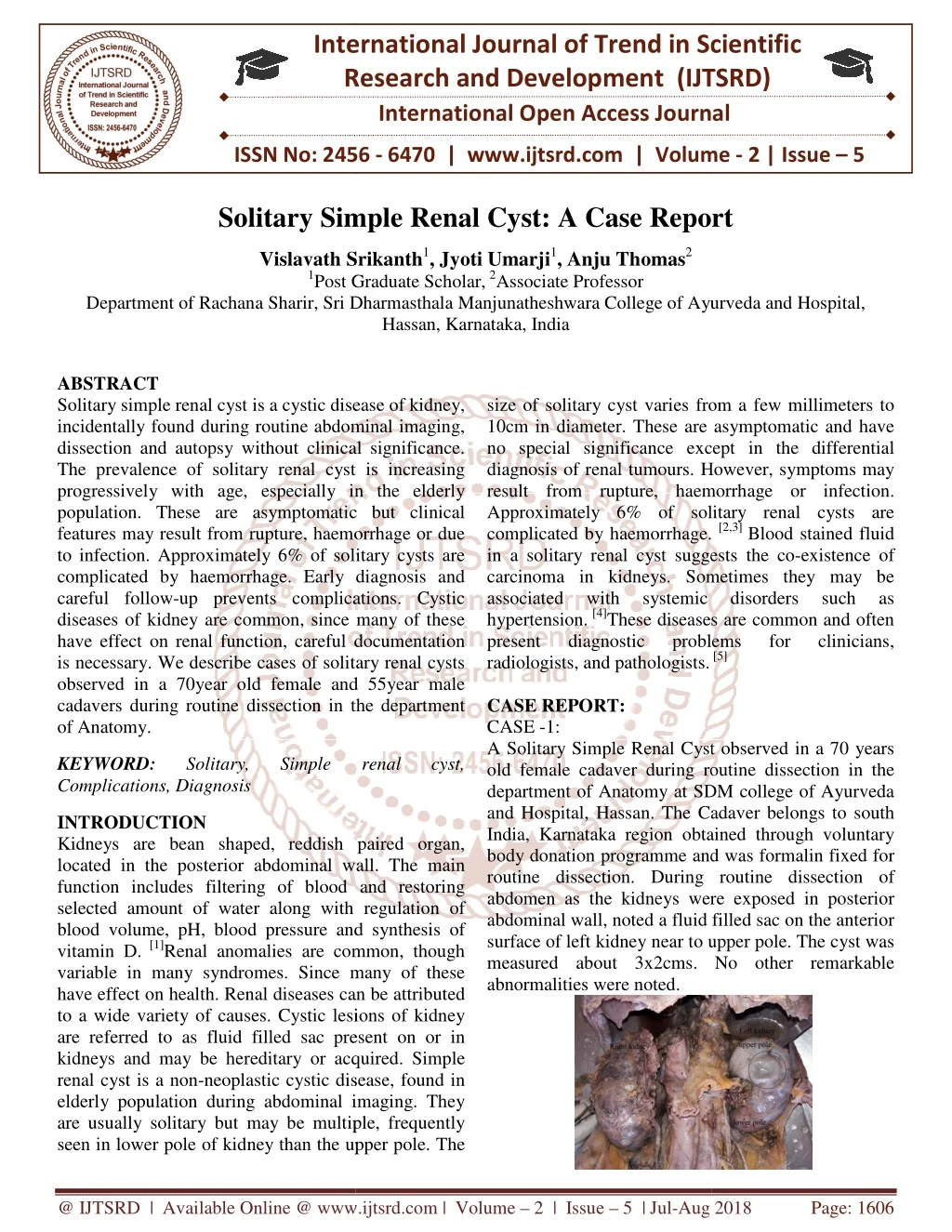
International Research Research and Development (IJTSRD) International Open Access Journal Solitary Simple Renal Cyst: A Case Report Vislavath Srikanth1, Jyoti Umarji1, Anju Thomas2 Post Graduate Scholar,2Associate Professor f Rachana Sharir, Sri Dharmasthala Manjunatheshwara College of Ayurveda Hassan, Karnataka, India International Journal of Trend in Scientific Scientific (IJTSRD) International Open Access Journal ISSN No: 2456 ISSN No: 2456 - 6470 | www.ijtsrd.com | Volume 6470 | www.ijtsrd.com | Volume - 2 | Issue – 5 Solitary Simple Renal Cyst: A Case Report Solitary Simple Renal Cyst: A Case Report Vislavath Srikanth 1Post Graduate Scholar Department of Rachana Sharir, Sri Dharmasthala Manjunatheshwara College f Ayurveda and Hospital, ABSTRACT Solitary simple renal cyst is a cystic disease of kidney, incidentally found during routine abdominal imaging, dissection and autopsy without clinical significance. The prevalence of solitary renal cyst is increasing progressively with age, especially in the elderly population. These are asymptomatic but clinical features may result from rupture, haemorrhage or due to infection. Approximately 6% of solitary cysts are complicated by haemorrhage. Early diagnosis and careful follow-up prevents complications. Cystic diseases of kidney are common, since many of these have effect on renal function, careful documentation is necessary. We describe cases of solitary renal cysts observed in a 70year old female and 55year male cadavers during routine dissection in the department of Anatomy. size of solitary cyst varies from a few millimeters to 10cm in diameter. These are asymptomatic and have no special significance except in the differential diagnosis of renal tumours. However, symptoms may result from rupture, haemorrhage or infection. Approximately 6% of solitary renal cysts are complicated by haemorrhage. in a solitary renal cyst suggests the co carcinoma in kidneys. Sometimes they may be associated with systemic hypertension. [4]These diseases are common and often present diagnostic problems radiologists, and pathologists. CASE REPORT: CASE -1: A Solitary Simple Renal Cyst observed in a 70 years old female cadaver during routine dissection in the department of Anatomy at SDM college of Ayurveda and Hospital, Hassan. The Cadaver belongs to south India, Karnataka region obtained through voluntary body donation programme and was formalin fixed for routine dissection. During routine dissection of abdomen as the kidneys were exposed in posterior abdominal wall, noted a fluid filled sac on the anterior surface of left kidney near to upper pole. The cyst was measured about 3x2cms. No other remarkable abnormalities were noted. cystic disease of kidney, ize of solitary cyst varies from a few millimeters to 10cm in diameter. These are asymptomatic and have no special significance except in the differential diagnosis of renal tumours. However, symptoms may result from rupture, haemorrhage or infection. oximately 6% of solitary renal cysts are complicated by haemorrhage. [2,3] Blood stained fluid in a solitary renal cyst suggests the co-existence of carcinoma in kidneys. Sometimes they may be associated with systemic se diseases are common and often present diagnostic problems incidentally found during routine abdominal imaging, dissection and autopsy without clinical significance. The prevalence of solitary renal cyst is increasing progressively with age, especially in the elderly asymptomatic but clinical features may result from rupture, haemorrhage or due to infection. Approximately 6% of solitary cysts are complicated by haemorrhage. Early diagnosis and up prevents complications. Cystic disorders disorders such such as as mon, since many of these have effect on renal function, careful documentation is necessary. We describe cases of solitary renal cysts observed in a 70year old female and 55year male cadavers during routine dissection in the department for for clinicians, clinicians, [5] A Solitary Simple Renal Cyst observed in a 70 years old female cadaver during routine dissection in the Anatomy at SDM college of Ayurveda and Hospital, Hassan. The Cadaver belongs to south India, Karnataka region obtained through voluntary body donation programme and was formalin fixed for routine dissection. During routine dissection of eys were exposed in posterior abdominal wall, noted a fluid filled sac on the anterior surface of left kidney near to upper pole. The cyst was measured about 3x2cms. No other remarkable KEYWORD: Complications, Diagnosis Solitary, Solitary, Simple Simple renal renal cyst, cyst, INTRODUCTION Kidneys are bean shaped, reddish paired organ, located in the posterior abdominal wall. The main function includes filtering of blood and restoring selected amount of water along with reg blood volume, pH, blood pressure and synthesis of vitamin D. [1]Renal anomalies are common, though variable in many syndromes. Since many of these have effect on health. Renal diseases can be attributed to a wide variety of causes. Cystic lesions of kidney are referred to as fluid filled sac present on or in kidneys and may be hereditary or acquired. Simple renal cyst is a non-neoplastic cystic disease, found in elderly population during abdominal imaging. They are usually solitary but may be multiple, frequently seen in lower pole of kidney than the upper pole. The seen in lower pole of kidney than the upper pole. The Kidneys are bean shaped, reddish paired organ, located in the posterior abdominal wall. The main function includes filtering of blood and restoring selected amount of water along with regulation of blood volume, pH, blood pressure and synthesis of Renal anomalies are common, though variable in many syndromes. Since many of these have effect on health. Renal diseases can be attributed to a wide variety of causes. Cystic lesions of kidney are referred to as fluid filled sac present on or in d may be hereditary or acquired. Simple neoplastic cystic disease, found in elderly population during abdominal imaging. They are usually solitary but may be multiple, frequently @ IJTSRD | Available Online @ www.ijtsrd.com @ IJTSRD | Available Online @ www.ijtsrd.com | Volume – 2 | Issue – 5 | Jul-Aug 2018 Aug 2018 Page: 1606
International Journal of Trend in Scientific Research and Development (IJTSRD) ISSN: 2456-6470 The prevalence of cyst is increasing progressively with age, though rare in children. [10]It is estimated that 25% people of 40 years and 50% people of 50 years have simple renal cysts. [11]The occurrence of cyst relatively equal in both gender, but C.C. Chang, et.al (2007) reported that the incidence was higher in males compared to females. [12]In present cases, the location and size of solitary cyst was variant. In the first case solitary cyst was located near the upper pole of the kidney, which was uncommon. The size was bigger may be due to early development and progressed with age. In second case cyst was at lower pole and small in size. It suggests that location of solitary cyst was uncertain and the size increases with age. The exact a etiology of cyst is not known. Some theories were proposed to justify the cause. It is thought to originate from diverticulae in the distal convoluted tubule, but this concept is disproved. [13] The accepted hypothesis is that segmental ischemia of kidney and an intra-renal calyceal obstruction are considered to be main factors in pathogenesis. [14] Morphologically, individual solitary simple renal cyst is oval to round in shape and size varies from a few millimetres to 10cm in diameter. [15]The cyst size increases in approximately one fourth of cases with age and an estimated rate is 1.6mm per year. It may double the original size over 10 years. [16]Solitary simple renal cysts are unilateral, filled with serous fluid enclosed by sac and present in the surrounding renal parenchyma. They are confined to the cortex and lined by single layer of cuboidal or flattened cuboidal epithelium. The characteristically yellowish-white and translucent. The cyst usually contains clear straw-coloured fluid, it may become rust-coloured due to haemorrhage. [17] Numerous classifications of renal cysts are proposed to get reasonable clinico-pathological correlation based on morphological patterns, radiographic appearance and vasculature, genetic analysis, and clinical evaluation of renal function. Most of classifications distinguish general strategies, which are continued till today. [18] Bosniak (1986) introduced new classification system morphological appearance and enhancement of renal cysts on CT and revised in 2003. The Updated Bosniak renal cyst classification was accepted by urologists and radiologists for the diagnosis and management of cystic lesions shown in Table.1.[19] Figure1: solitary renal cyst near upper pole of left kidney. CASE 02: This is a case of Solitary renal cyst observed in 55year old male cadaver during routine dissection in the department of Anatomy at SDM college of Ayurveda and Hospital, Hassan. The cyst was located at lower pole of right kidney. Measurement of the cyst was performed directly and measured as 0.5x0.2cm. No other remarkable abnormalities were noted wall of cyst is Figure 2:Solitary cyst on lower pole of the Right kidney DISCUSSION: Cyst is a fluid filled sac, it may be true cyst or false. True cyst contains serous or mucoid fluid and wall of sac is lined by epithelial layer. The fluid accumulation is due to secretion of lining epithelium. The false one does not have any epithelial lining. [6] Renal cyst is defined as presence of liquid or semisolid fluid in an enclosed sac in or on the kidneys. Cystic diseases of kidney are hereditary, developmental or acquired. Simple renal cyst is a non-neoplastic cystic kidney disease, presented unilaterally. These are incidentally found during routine abdominal imaging, dissection and autopsy studies. They are usually solitary but may be multiple, frequently seen in lower pole of kidney than the upper pole. [7,8] Simple renal cyst may be present at birth but resolved after birth. [9] depending on the @ IJTSRD | Available Online @ www.ijtsrd.com | Volume – 2 | Issue – 5 | Jul-Aug 2018 Page: 1607
International Journal of Trend in Scientific Research and Development (IJTSRD) ISSN: 2456 International Journal of Trend in Scientific Research and Development (IJTSRD) ISSN: 2456 Table.1.The updated Bosniak renal cyst classification. Septa International Journal of Trend in Scientific Research and Development (IJTSRD) ISSN: 2456-6470 Table.1.The updated Bosniak renal cyst classification. Septa Calcification Stage Cyst wall Enhancement Management No follow-up No follow-up CT: 3, 6, 12 monthly then annual I Hairline thin No No No No Minimal regular thickening Minimal regular thickening Irregular thickening Gross, irregular thickening Smooth, hairline thin II Few,hairline thin Few,hairline thin No Multiple, minimal smooth thickening Measurably thick, irregular Irregular gross thickening thickening up. Cyst size of diameter of 3 cm is also an indication for follow-up. : indeterminate Stage III should be managed as IIF, while definitive Stage III should be managed surgically. : indeterminate Stage III should be managed as IIF, while definitive Stage III should be managed surgically. Multiple, minimal smooth thickening Measurably thick, irregular Irregular gross IIFa Thick, nodular No Thick, nodular, irregular Thick, nodular, irregular IIIb Yes As IIF or surgical yes, tissue and cyst IV Surgical IIFa : Denotes follow-up. Cyst size of diameter of 3 cm is also an indication for follow IIIb: indeterminate Stage III should be managed as IIF, while definitive Stage III should be managed surgically. The above classification will give an idea of different cases into non-surgical (category I and II), and surgical (category III and IV). Solitary simple renal cysts are usually asymptomatic. In some cases, however, pain may occur between the ribs and hips, when cyst enlarges and press on other underlying structures. [20]The large cyst may appear as lump in loin or back. If patient complaints pain in loin with malaise and fever, it suggests infected cyst. Severe loin pain with mild haematuria symptoms may develop from haemorrhagic cyst. Cyst on hilum of kidney or pelviureteric junction causes urinary symptoms. A large solitary cyst may produce renal insufficiency and uraemia by damaging renal tissue. [21]Solitary cyst do not affect the renal function, but one study found an association between the presence of cyst and reduced renal function in hospitalized people younger than 60 years of age. [22] (2013) found a positive relationship between solitary renal cysts and prehypertension as well as hypertension. [23] A study by Pedersen et al (1993) showed that mean arterial blood pressure was significantly higher in individuals with simple renal cysts. [24] (2009) reported that aspiration of cysts resulted reduction of hypertension. However, this relationship is not well understood. [25] N.Terada et al (2004)reported old age, male gender, renal dysfunction, and hypertension were considered to be risk factors for solitary renal cysts. Chang et al(2007)expressed same as above and also found renal stones, serum creatinine, and smoking are other risk factors.[27]Approximately 6% of simple cysts are complicated by haemorrhage. Blood stained cysts are complicated by haemorrhage. Blood stained up. The above classification will give an idea of different surgical (category I and II), and fluid in a solitary cyst suggests towards the co- of carcinoma. Occasionally, renal adenoma or papillary adenocarcinoma arises in the walls of the fluid in a solitary cyst suggests towards the co existence of carcinoma. Occasionally, renal adenoma or papillary adenocarcinoma arises in the walls of the simple renal cysts. [28] The accurate diagnosis helps in differentiating renal tumours and appropriate management. Straight x may show some portion of kid streaks of calcium on the cyst wall. The Excretory urography will show displacement of upper part of ureter, when a large [29]Ultrasonography defines a simple cyst with well transmitted sound waves and absence of any Any complexity that deviates from this should be further evaluated by CT. Computed Tomography helps in accurate diagnosis along with differentiating renal cyst from tumour. It confirms complexity of the cyst with presence of calcification, septae, wall thickening or nodularity and helps to evaluate the associated risk for malignancy. Magnetic resonance imaging has better contrast resolution than CT. It is useful to determine type II and III stage of cyst, but not necessary for routine evaluation. Solitary renal cysts are asymptomatic, until unless infected or ruptured. [31]Symptomatic cysts can be managed with a variety of surgical and percutaneous methods including percutaneous aspiration (with or without a sclerosing marsupialization, open unroofing. The Simple drainage without sclerotherapy is associated with recurrence; by using a sclerotic agent provides more satisfactory results than aspiration alone. Percutaneous drainage with single multiple-session sclerotherapy has been successfully performed with high success rates. Several authors reported, multiple-session sclerotherapy has better session sclerotherapy has better Solitary simple renal cysts are usually asymptomatic. occur between the The accurate diagnosis helps in differentiating renal tumours and appropriate management. Straight x-ray may show some portion of kidney expansion with streaks of calcium on the cyst wall. The Excretory urography will show displacement of upper part of ureter, when a large Ultrasonography defines a simple cyst with well- transmitted sound waves and absence of any echoes. Any complexity that deviates from this should be further evaluated by CT. Computed Tomography helps in accurate diagnosis along with differentiating renal cyst from tumour. It confirms complexity of the cyst with presence of calcification, septae, loculation, wall thickening or nodularity and helps to evaluate the associated risk for malignancy. Magnetic resonance imaging has better contrast resolution than CT. It is useful to determine type II and III stage of cyst, but ribs and hips, when cyst enlarges and press on other The large cyst may appear as lump in loin or back. If patient complaints pain in loin with malaise and fever, it suggests infected cyst. h mild haematuria symptoms may develop from haemorrhagic cyst. Cyst on hilum of kidney or pelviureteric junction causes urinary symptoms. A large solitary cyst may produce renal insufficiency and uraemia by damaging renal tissue. ffect the renal function, but one study found an association between the presence of cyst and reduced renal function in hospitalized pole. pole. cyst cyst at at lower lower [22] C.T. Lee et al (2013) found a positive relationship between solitary and prehypertension as well as aluation. [30] A study by Pedersen et al (1993) showed that mean arterial blood pressure was significantly higher in Solitary renal cysts are asymptomatic, until unless Symptomatic cysts can be managed with a variety of surgical and percutaneous methods including percutaneous aspiration (with or without a sclerosing marsupialization, open unroofing. The Simple drainage without sclerotherapy is associated with recurrence; by using a sclerotic agent provides more satisfactory results than Percutaneous drainage with single or session sclerotherapy has been successfully performed with high success rates. Several authors [24]Zerem et al (2009) reported that aspiration of cysts resulted in a reduction of hypertension. However, this relationship agent), and and agent), percu percutaneous laparoscopic laparoscopic cyst cyst N.Terada et al (2004)reported old age, male gender, renal dysfunction, and hypertension were considered to be risk factors for solitary renal cysts.[26]C.C. l(2007)expressed same as above and also found renal stones, serum creatinine, and smoking are Approximately 6% of simple @ IJTSRD | Available Online @ www.ijtsrd.com Available Online @ www.ijtsrd.com | Volume – 2 | Issue – 5 | Jul-Aug 2018 Aug 2018 Page: 1608
International Journal of Trend in Scientific Research and Development (IJTSRD) ISSN: 2456-6470 results in preventing the reoccurrence of cyst. [32]Kirwin’s operation (a portion of cyst wall removed and cavity is filled with perinephric fat then suture with corrugated drain) is curative method in which reoccurrence is impossible33.Occasionally, the complicated solitary cyst located on one of the pole needs partial nephrectomy. [34] CONCLUSION: Simple renal cyst is a cystic disease of kidney, incidentally found during routine abdominal imaging, dissection and autopsy with no clinical significance. The prevalence and size increases progressively with advancing age. The presence of solitary renal cysts in a potential kidney donor is not a barrier to renal transplantation but long-term follow up is needed. An awareness of such a presence is valuable for the surgeons and radiologists in differential diagnosis of other cystic diseases of kidney and Renal tumours. REFERENCE: 1.Tortora GJ, Grabowski SR, Principles of Anatomy and Physiology, 18thed, New York: HarperCollins College; 1996, p 849. 2.Mohan H, Text Book of Pathology, 4thed, New Delhi: Jaypee Brothers medical publishers (PVT) Limited; 2010, p644. 3.Kumar P, Clark M. Kumar & Clark's Clinical Medicine, 7thed, Edinburgh: Saunders/Elsevier; 2009, p642. 4.Chatterjee BP, A Short Textbook of Surgery, 3rded, Vol.1. Calcutta: New Central Book Agency (p) Limited; 1993, p345-346. 5.Koh C, et al. The management of symptomatic simple renal cysts in children, J PedSurg Case Reports; 2018.28: 21-29. 6.Das. S, A Concise Textbook of Surgery, 6thed, Kolkata: Dr. S. Das publication; 2010,p 94-95. 7.Mohan H, Text Book of Pathology, 4thed, New delhi: Jaypee Brothers medical publishers (PVT) Limited; 2010, p640-644. 8.Kumar P, Clark M. Kumar & Clark's Clinical Medicine, 7thed, Edinburgh: Saunders/Elsevier; 2009, p642. 9.Zerem E, Imamovic G, Omerovic S, Simple renal cysts and arterial hypertension: does their evacuation decrease the blood pressure?, Journal of Hypertension; 2009, 27:2074–2078. 10.Simms RJ, Ong ACM, How simple are ‘simple renal cysts’? Nephrol Dial Transplant; 2014, 29: iv106-112. 11.Torres VE, Grantham JJ, Brenner & Rector’s The Kidney, 8thed, Vol. 2. Philadelphia: Saunders Elsevier; 2008, 1451–1453. 12.C. C. Chang, et al. Prevalence and Clinical Characteristics of Simple Renal Cyst, J Chin Med Assoc; 2007, 70(11):486–491. 13.Simms RJ, Ong ACM, How simple are ‘simple renal cysts’? Nephrol Dial Transplant; 2014, 29: iv106-112. 14.Chatterjee BP, A Short Textbook of Surgery, 3rded, Vol.1. Calcutta: New Central Book Agency (p) Limited; 1993, p 345-346. 15.Mohan H, Text Book of Pathology, 4thed, New Delhi: Jaypee Brothers medical publishers (PVT) Limited; 2010, p640-644. 16.Eknoyan GA, Clinical view of simple and complex renal cysts, J Am SocNephrol; 2009, 20:1874–1876.28. 17.Mohan H, Text Book of Pathology, 4thed, New Delhi: Jaypee Brothers medical publishers (PVT) Limited; 2010, p640-644. 18.Bonsib SM, The Classification of Renal Cystic Diseases and Other Congenital Malformations of the Kidney and Urinary Tract, Arch Pathol Lab Med;2010, 134:554-567. 19.Eknoyan GA, Clinical view of simple and complex renal cysts, J Am SocNephrol; 2009, 20:1874–1876.28. 20.www.kidney.niddk.nih.govt. Information /Kidney Cysts). 21.Das. S, A Concise Textbook of Surgery, 6thed, Kolkata: Dr S. Das publication; 2010, p1038- 1039. 22.Torres VE, Grantham JJ, Brenner & Rector’s The Kidney, 8thed, Vol. 2. Philadelphia: Saunders Elsevier; 2008, 1451–1453. 23.C-T Lee, et al. Multiple and large simple renal cysts are associated with prehypertension and hypertension, Kidney International; 2013, 83:924– 930. (Home /Health Kidney Disease /Simple @ IJTSRD | Available Online @ www.ijtsrd.com | Volume – 2 | Issue – 5 | Jul-Aug 2018 Page: 1609
International Journal of Trend in Scientific Research and Development (IJTSRD) ISSN: 2456-6470 24.C.C. Chang, et al. Prevalence and Clinical Characteristics of Simple Renal Cyst, J Chin Med Assoc; 2007, 70(11):486–491. 25.Zerem, et al. Simple renal cysts and arterial hypertension: does their evacuation decrease the blood pressure, Journal of Hypertension; 2009, 27(10):2074–2078. 26.Terada N, et al. Risk factors for renal cysts, B J U International; 2004, 93:1300–1302 27.C.C. Chang, et al. Prevalence and Clinical Characteristics of Simple Renal Cyst, J Chin Med Assoc; 2007, 70(11):486–491. 28.KumarV, Robbins Basic Pathology, 9thed, USA: Elsevier India (PVT) Limited, reprint2014, p542. 29.Das. S, A Concise Textbook of Surgery, 6thed, Kolkata: Dr S. Das publication; 2010, p1038- 1039. 30.Eknoyan GA, Clinical view of simple and complex renal cysts, J Am SocNephrol; 2009, 20:1874–1876.28. 31.Kumar P, Clark M. Kumar & Clark's clinical medicine, 7thed, Edinburgh: Saunders/Elsevier; 2009, p642. 32.Akinci D, et al. Long-term results of single- session percutaneous drainage and ethanol sclerotherapy in simple renal cysts, European Journal of Radiology; 2005, 54:298–302. 33.Das. S, A Concise Textbook of Surgery, 6thed, Kolkata: Dr S. Das publication; 2010, p1038- 1039. 34.Bhat SM, SRB’s Surgical Operations: Text and Atlas in: urologic surgeries, 1sted, New Delhi: Jaypee publications; 2014, p1085. @ IJTSRD | Available Online @ www.ijtsrd.com | Volume – 2 | Issue – 5 | Jul-Aug 2018 Page: 1610