Download
1 / 17
170 likes | 335 Vues
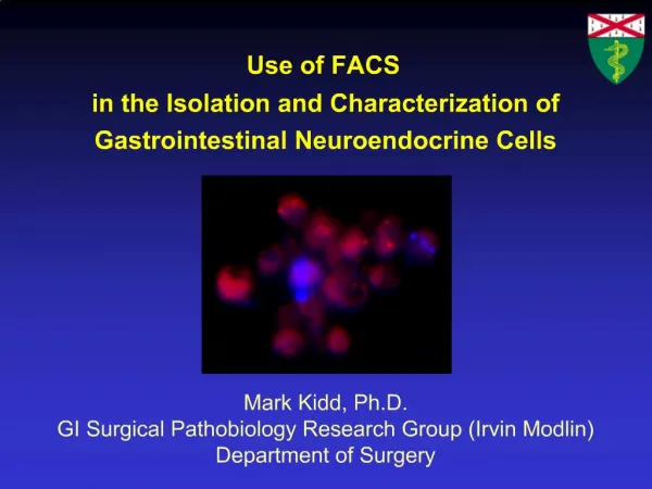
Use of FACS in the Isolation and Characterization of Gastrointestinal Neuroendocrine Cells. Mark Kidd, Ph.D. GI Surgical Pathobiology Research Group (Irvin Modlin) Department of Surgery. Gastrointestinal Neuroendocrine cell Neoplasia “Carcinoid”.

E N D
Use of FACS in the Isolation and Characterization of Gastrointestinal Neuroendocrine Cells Mark Kidd, Ph.D. GI Surgical Pathobiology Research Group (Irvin Modlin) Department of Surgery
Gastrointestinal Neuroendocrine cell Neoplasia “Carcinoid” • Tumor incidence has increased 700-2700% since 1983* • Little is known about the physiology or pathobiology • No significant advances in therapeutic modalities • Neuroendocrine cells = Progenitor cells of neoplasia • No pure naïve neuroendocrine cell preparation *NCI (1973-2002)
Gastrointestinal Neuroendocrine Cells • 1-2% by volume of mucosa • Sequestrated in crypts within the mucosa Difficult cells to isolate and examine
Previous protocols for neuroendocrine cell isolation • Mucosal scrapping or inverted mucosal sacs • Digestion with pronase/collagenase • Respiration/calcium-free media = Cell slurry ~1-2% pure neuroendocrine cells • Nycodenz gradient centrifugation • Elutriation • Short-term culture 50-70% 72-84% 80-90% Enrichment Significant enrichment but not homogeneous
Characteristics potentially useful for FACS • Size • Density • Acidic vesicles • Vesicular monoamine transporters (VMAT) • Acid gradient • Accumulates weak bases [Amine] [Amine] VMAT 1/2 [H+] V-type ATPase [H+] pH Vesicles accumulate weak bases
Acridine Orange Nuclei →fluoresce green Cytoplasmic RNA→fluoresce orange Absorption Emission FITC/Cy7 channel AO widely used as a pH-sensitive dye in studies of acid secretion
Acridine Orange Parietal cells Neuroendocrine cells pH 1-2 pH 3-5 AO Accumulation Stacking AO Accumulation No stacking pH determines emission
Acridine Orange – Gastric mucosa FACS Parietal cells* Orange/Red Neuroendocrine ECL cells* *95-99% pure Lambrecht N et al. Physiol Genomics 2006; 25:153-65 Green Rodent gastric cell populations separated by AO fluorescence
ECL Results – Human Gastric ECL cells 97.3-99.1% pure (HDC-positive) • 92.9-95.6% viable • Proliferate in short-term culture Human gastric neuroendocrine ECL cells separated by AO fluorescence
Protocol developed for Small Intestinal EC cells FACS approach Mixed cell population used for control studies Terminal ileum Mixed cell population F0 (~4%EC cells) Collagenase/pronase digestion of tissue at 37OC for 1 hour Nycodenz gradient centrifugation FN (~75% pure EC cells) 1.07 g/l Immunostaining of FN with acridine orange 99% Pure live EC cell preparation ~ 1 million cells Confirm by EM/confocal microscopy, immunostaining and PCR of neuroendocrine markers, measure serotonin content FACS of live EC cells Kidd M. et al. Am J Physiol Gastrointest Liver Physiol. 2006 Feb 2; [Epub ahead of print]
FACS sorting – Human Small Intestinal Mucosa [AO] =50-200nM 1% of nuclear stain
Results – Human Small Intestinal EC cells A B 99% preparations of naïve human EC cells Modlin I.M. et al. J Clin Endocrinol Metab. 2006 Mar 14; [Epub ahead of print]
Secretion – Human Small Intestinal EC cells Isoproterenol Forskolin EC50 = 8.1x10⁻⁸M EC50 = 2.1x10⁻⁷M Short-term culture Serotonin secretion cAMP/adrenergic control
Summary • Method established for gastric ECL cells • Small intestinal EC cells can be isolated by similar approach • Viable, highly purified preparations • Short-term culture • Proliferation/secretory studies • Transcriptome analysis • Define cellular regulators = Understand physiology • = Unravel pathobiology • = Identify new therapeutic targets Future Directions
Acknowledgements Irvin Modlin GI Pathobiology Research Group Dept. Surgery, Yale Pathology, Yale Keck, Yale FACS, Yale Physiology, UCLA Manish Champaneria Geeta Eick Robert Camp Shrikant Mane Geoff Lyon Mark Shlomchik George Sachs Nils Lambrecht