Download
1 / 44
580 likes | 1.76k Vues
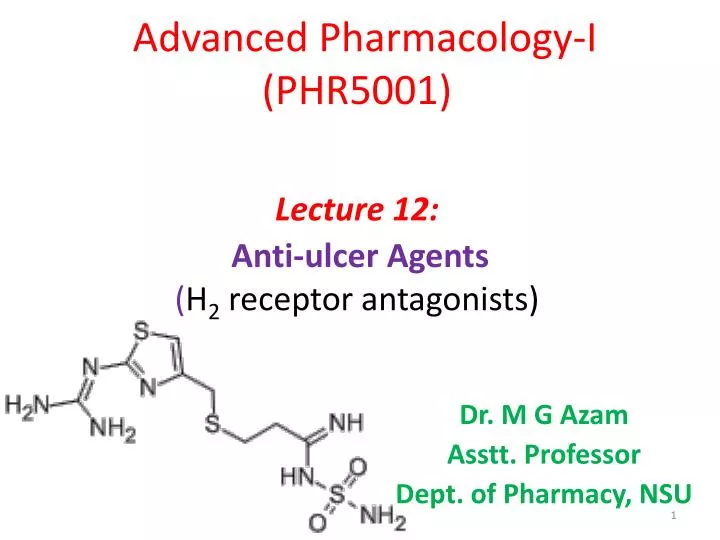
Advanced Pharmacology-I (PHR5001) Lecture 12: Anti-ulcer Agents ( H 2 receptor antagonists ). Dr. M G Azam Asstt . Professor Dept. of Pharmacy, NSU. Introduction. Definition: Anti-ulcer ( Antisecretory ) agents are the drugs which decreases the secretion of gastric acid in the stomach.

E N D
Advanced Pharmacology-I(PHR5001)Lecture 12:Anti-ulcer Agents(H2 receptor antagonists) Dr. M G Azam Asstt. Professor Dept. of Pharmacy, NSU
Introduction Definition: Anti-ulcer(Antisecretory) agents are the drugs which decreases the secretion of gastric acid in the stomach. • Histamine - Powerful stimulant of Hydrochloric acid secretion from parietal (oxyntic)cells of gastric mucosa. • Histamine (In larger dose )- increase secretion of pepsin. These actions are mediated by H2-receptor. • The secretion of acid by these parietal cells are regulated by various mediators or receptors. • Histamine H2 receptors • G receptors-release gastrin & release gastric acid • Acetylcholine (Ach) M3receptor
What is Peptic Ulcer ? • A peptic ulcer disease or PUD is an ulcer (defined as mucosal erosions ≥ 0.5 cm) of an area of the gastrointestinal tract exposed to the acid and pepsin secretion • Gastritis is the precursor to PUD and it is clinically difficult to differentiate the two • Stomach (called gastric ulcer): Relieved by food but pain may persist even after eating. Infrequent or absent remissions (>55 yrs) • Duodenum (called duodenal ulcer): burning upper abdomen pain relieved by food but reappears 1-3 hrs after meals. Worse pain when stomach empty • Esophagus (called Esophageal ulcer) Causes of ulcers: • Hypersecretion of acid and pepsin and GI infection of gm-ve bacteria H. Pylori. • Stress, alcohol, ulcerogenic drugs (e.g. NSAIDs), male genders, age, & diet
Why Ulceration Occurs? • High [H+] in the gastric lumen • Require defense mechanisms to protect oesophagus and stomach • Mucus secretion: slows ion diffusion • Prostaglandins: I2 and E2 (alcohol, aspirin ) • Bicabonate ions • High Blood Flow (nitric oxide) • Imbalance primarily between Aggressive factors • & Defensive factors: Defensive factors, e.g. mucus, HCO3, PG Aggressive factors, e,g, acid, pepsin, bile etc.
Gastroesophageal Reflux Disease (GERD) • Common and GI motility disorder • Acidity of Gastric contents – most common factor • Acid contents reflux back into esophagus • Intense burning, sometimes belching • Can lead to esophagitis, and esophageal ulcers • Barrett’s esophagus (A complication of severe chronic GERD involving changes in the cells of the tissue that line the bottom of the esophagus. These esophageal cells become irritated when the contents of the stomach back up (refluxes) and there is a small but definite increased risk of adenocarcinoma of the esophagus.) • Commonly associated with obesity • Improves with lifestyle management
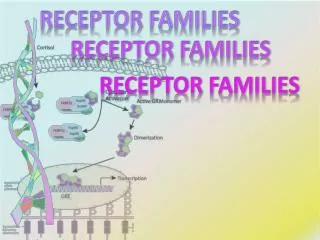
Physiology of Acid Secretion Schematic diagram the physiologic control of hydrogen ion secretion by the gastric parietal cell. ECL (enterochromaffin-like) cell; G(CCK-B), gastrin-cholecystokinin-B receptor; H, histamin; M1, M3, muscarinic receptors; ST2, somatostatin-2 receptor; ATPase, H+/K+ ATPase proton pump.
Histamine uses the CAMP pathway whereas gastrin and Ach uses Ca2+ dependent pathway. Both pathway activates the H+/K+ ATPase. Ach release from postganglionic vagal fibres can stimulate directly gastric acid secretion though M3 (a specific muscarnic cholinergic receptor subtype). Ach. also indirectly affects the parietal cells though the stimulation of histamine release from the entrochromaffin-like cells in the fundus and the stimulation of gastric release from G cells in the gastric antrum. • Histamine is released from ECL cells through the multifactorial pathway and is critical regulator of acid production through the H2 sub type of receptor. • The release of gastric is regulated through the multifactorial pathway and it stimulates acid secretion predominantly in an indirect manner by causing the release of histamine from ECL cells; a direct effect of gastric or parietal cell is also seen but is less important.
H2 receptor antagonist Comparison H1 receptor antagonist Two aryl, heteroaryl rings in place of imidazole (bulky groups) Connecting chain of aryl and terminal nitrogen is of 2 to 3 atoms Ionic flexible chain at end. Ionized at pH 7.4 Hydrophobic (due to aryl rings). High partition coefficient value Low dipole moment Imidazole or other 5 membered heterocyclic ring (Imidazole is not necessary) Connecting chain of ring and terminal nitrogen is of 4 atoms Polar π e containing systems, amphoteric, unionized at pH 7.4 Hydrophobic (due to polar grp). Low partition coefficient High dipole moment
Treatment of ulcer • H2 receptor antagonists are the most popular drug for the treatment of peptic ulcer. H2 receptor antagonists Must bind but not activate H2 receptor site • They bring about sympathomimetic relief and promote ulcer healing. • Gastric acid secretion involve H2 receptor which is competitively blocked by H2 blockers. • In 1972, Black and co-workers first described selective H2 receptor blokade for acid secretion. With successful introduction of cimetidine, in 1977 other analogs like ranitidine, famotidine & roxatidine has been synthesized.
Imidazole ring replaced by various Nitrogen containing heterocyclic ring to optimize the activity. Furan (Ranitidine) & thiazole (Nizatidine) showed good activity. • Substitution in side chain with different basic as well as neutral groups were carried out like nitroamine ketene (Ranitidine) & sulfonylamidine(Famotidine) for better activity. • Substitution of dimethyl amino methyl (Ranitidine & Nizatidine) and amidine • (Famotidine) groupson ring showed better activity.
Classification of Anti-ulcer Drugs • Gastric Antisecretory Drugs HCl secretion • Antagonize Rs. on Parietal Cell--- H2,M3, G • Inhibitor of H+-Pump II. Antacids---Neutralizations of secreted acid and pepsin activity III. Agents killHP : Eradication of Helicobacter pylori by triple therapy: Almost all duodenal and 2/3 gastric ulcer pt’s infected with HP Omeprazole / Lansoprazole - 20 / 30 mg bd Clarithromycin - 500 mg bd Amoxycillin / Metronidazole - 1gm / 500 mg bd Given for 14 days
HCl & pepsin Misoprostol Ranitidine + _ _ _ _ _ + + + + + + + Omeprazole Antacid • Probanthine • Pirenzepine Gastrin PGE2 Histamine Proglumide ACh • Compete Gastrin-R H2 M3 Adenyl cyclase Gastrin receptor PGE receptor Ca++ ATP cAMP Ca++ Protein Kinase (Activated) K+ H+ K Parietal cell Proton pump Lumen of stomach Gastric acid
Classification of Anti-ulcer Drugs • Reduction in Gastric acid secretion: • H2 antihistamines: Cimetidine, Ranitidine, Famotidine, Nizatidine and Roxatidine • Proton pump inhibitors: Omeprazole, Lansoprazole Pantoprazole, Rabeprazole and Esomeprazole • Anticholinergics:Pirenzepine, Propantheline and Oxyphenonium • Prostaglandin analogue: Misoprostol • 2. Acid Neutralizing agents: (ANTACIDS) • Systemic: Sodium Bicarbonate and Sod. Citrate • Nonsystemic: Magnesium hydroxide, Mag. Treisilicate, Aluminium hydroxide gel, Magaldrate and calcium carbonate Ulcer protectives: Sucralfate, Colloidal Bismuth sudcitrate Anti-H. pylori Drugs: Amoxicillin, Clarithromycin, metronidazole, tinidazole and tetracycline
H2 Antagonists • Cimetidine, Ranitidine, Famotidine, Roxatidine, Nizatidine • MOA: • Reversible competitive inhibitors of H2 receptor • Highly selective, no action on H1 or H3 receptors • All phases of gastric acid secretion • Very effective in inhibiting nocturnal acid secretion (as it depends largely on Histamine ) • Modest impact on meal stimulated acid secretion (as it depends on gastrin, acetylcholine and histamine) • Volume of pepsin content and IF are also reduced • Volume reduced by 60 – 70% - anti ulcerogenic effect • Adverse Effect: • Gynecomastia, prolactin , CYP450 , headache
Proton Pump Inhibitors Omeprazole • Most effective drugs in antiulcer therapy • Prodrugsrequiring activation in acidic pH. • Block enzymes responsible for secreting HCl - binds irreversibly to H+K+ATPase • Prototype: Omeprazole • Substituted Benzimidazole derivative • Diffuses into G. canaliculi = accumulation pH < 5 (proton catalyzed )= tetracyclic sulfenamide + sulphenic acid • Covalent binding with sulfhydryl cysteines of H⁺K⁺ ATPase • Irreversible inactivation of the pump molecule • Acid suppressants regardless of stimulating factors • Also inhibits gastric mucosal carbonic anhydrase
CLINICAL USES OF AGENTS AFFECTING GASTRIC ACIDITY: 1. H2 –histamine receptor antagonist eg. Ranitidine,Cimetidine,Famotidine, Nizatidine - Peptic ulcer (Gastric ulcer and duodenal ulcer) - Reflex oesophagitis 2. Proton Pump Inhibitor : Omeprazole Lansoprazole, Esomeprazole,Rabeprazole,Pantoprazole - Peptic ulcer - Reflex oesophagitis - As one component Of therapy for H-pylori infection - Zollinger-Ellison syndrome 3. Antacid eg. Mg-trisillicate, Al-hydroxide alginates - Dyspepsia - Symptomatic relief of peptic ulcer - Esophageal reflex 4. Bismuth chelate -One component of therapy for H-pylori infection.
Screeningmethods:- • Pyloric ligation induced gastric ulceration 2. Ethanol induced mucosal damage in rats 3. Stress ulcer through immobilization stress Lower & upper extremities fixed, wrapped in a wire gaze, kept at dark for 24 hr. 4.Indomethacin induced ulcers in rats After 10 min of admistering 20 mg/kg indomethacin, test drug is given orally. 6hr later , sacrifice in co2 , stomach is removed.
Pyloric ligation induced gastric ulceration • After 1h of treatment with test or std. (Ranitidine 50 mg/kg) , animals are anaesthetized with the help of anesthetic ether; the abdomen is opened by a small midline incision. Pyloric portion of the stomach is slightly lifted out and ligated. • The stomach is replaced carefully and the abdominal wall is closed by interrupted sutures. Rats are sacrificed by an over dose of anaesthetic ether after four hours of pyloric ligation. • Abdomen is opened and ligature is placed around esophagus. Stomach is removed and contents are drained to centrifuge tube. • The volume of the gastric juice is measured and centrifuged at 2000 rpm for 10 min. From the supernatant, aliquots (1 ml of each) are taken for the determination of pH, total and free acidity. Each stomach is examined (by a 10Χ magnifier lens) for lesions in the fore stomach portion & indexed according to severity. 0 - no ulcer, 1 - spot ulcer, 2 - deep ulcer, 3 - perforation % Inhibition of Ulceration =
Ethanol induced ulcer model The ulcer is induced by administering ethanol to Albino rats (fasted for 36 h). One group represented the control group, which received ethanol Second & Third • Groups received methanolic extract of A. Indicum 250 and • 500 mg/kg and, Ranitidine, in the dose of 50 mg/kg were • administered orally for Four group as reference standard • drug. The gastric ulcers were induced in rats by • administrating absolute ethanol (90%) (0.5 ml/100g) orally, • after 45 min of methanolic extract and ranitidine treatment. • They were kept in specially constructed cages to prevent • coprophagia during and after the experiment. The animals • were anaesthetized 1h latter with anaesthetic ether and • stomach was incised along the greater curvature and • ulceration will be scored
Advanced Pharmacology-I(PHR5001)Lecture 13:MONOCLONAL ANTIBODIES AS THERAPEUTIC AGENTS Dr. M G Azam Asstt. Professor Dept. of Pharmacy, NSU
Antibody is a class of immunological proteins (globular glycoproteins/ “Immunoglobulin” produced by plasma cells (B lymphocytes) in response to a specific antigen. • Antibodies function as markers, binding to the antigen so that the antigen molecules can be recognized and destroyed by phagocytes. • The part of the antigen that the antibody binds to is called the epitope. The epitope is thus a short amino acid sequence that the antibody is able to recognize. • Antibodies are useful tools for the immune system to combat pathogens.
MONOCLONAL ANTIBODIES (Mab) Polyclonal Abs • An antibody produced by a single clone of cells. monoclonal antibody is a single pure type of antibody. • MAbs are identical. They are directed toward same epitope. So more efficient and specific. • Mixture of antibodies similar to sera • Antibodies that are obtained from different B cell resources. They are a combination of immunoglobulin molecules secreted against a specific antigen, each identifying a different epitope. * Used in • Biomedical research • Diagnosis of diseases • Treatment of diseases such as infection, cancer, etc.
Structure of antibody • Consist of four polypeptide chains, somewhat resembling to a Y shape.i) 2 identical Light chains ( L chains) ii) 2 identical Heavy chains ( H chains) • Disulphide bonds hold 4 polypeptide chains Ig domain: 110 amino acids; globular domain used in many proteins. Variable domains, Constant domains, Hinge. Fab: fragment antigen binding Fc: fragment crystallizable (effector functions) CDR: Complementarity-Determining Regions
Production of Monoclonal Antibodies • A mouse is immunized with the specific antigen“X”. This will allow the mouse to produce antibodies. • The spleen cells producing specific antibodies are removed. • Fusion of spleen cells with tumor cells (myeloma cells) in a culture medium is done. The resulting cell is called a hybridoma. • Hybridoma cells are continuously growing cell line. Each hybridoma will produce relatively large quantities of monoclonal antibody molecules. These hybridomas have antibody producing capability from lymphocytes & have ability to grow continuously (immortal) from malignant cells. • Separate the hybridomas from unfused cells. • Culture selected hybridoma cells for the production of monoclonal antibodies in large quantity. • Remain indefinitely and maintained by in vitro; that is, in culture vessels or in vivo-in the body of an animal so that antibodies will be produced in body and can be recovered later from body fluid.
Purification of monoclonal antibody : • Affinity chromatography is used for purification of mAbs. • This method separates molecules on their differing affinities for a ligand. • Antigen can be bound to the support matrix (small, chemically reactive beads) in order to purify antigen-specific antibody from a polyclonal antiserum. • Specific antibody is eluted by altering the pH, which disrupt antibody-antigen bonds.
Application of monoclonal antibody : 1. Research tools: • Identification of cell surface marker • Purification of proteins and enzymes • Detection assays • Genetic engineering • Structure of cell membrane • Human: -umab • Humanized: -zumab • Murine: -momab • Chimeric: -ximab Sufix of Antibody 2. Clinical diagnosis: • Cancers • Leukemia • Viral disease • Allergic reaction • AIDS 3. Treatment of diseases:
Diagnostic tests Monoclonal antibodies are widely used as diagnostic and research reagents. • Once monoclonal antibodies for a given substance have been produced, they can be used to detect the presence of this substance. The Western blot test detect the protein on a membrane. • Monoclonal antibodies can also be used to purify a substance with techniques called affinity chromatography. • Other monoclonal antibodies allow rapid diagnosis of hepatitis, influenza, herpes and streptococcal infections. • Antibodies are used in several diagnostic tests to detect small amounts of drugs, toxins or hormones. • Monoclonal Antibodies also used in ELISA (enzyme linked immunosorbant Assay) for detection of viruses.
Monoclonal antibody in cancer therapy: • By means of genetic engineering, so-called chimeric antibodies are prepared that contain both human and mouse sections. • The mouse section is located in the variable region of the antibody that contains the antigen- binding site, while the human section is in the constant region. • After the variable region binds to the cancer antigen, the human section activates complement and cytotoxic T cells to destroy the cancer cell. • Naked mAbs:Without any drugs, radioactive materials or toxins. • B. Conjugated mAbs:They are combined with chemotherapy • drugs (ChemolabeledAb) or radioactive material or toxins (Immunotoxin)
When a monoclonal antibody attaches to a cancer cell, it can: • Make the cancer cell more visible to the immune system • Block growth signals • Deliver radiation to cancer cells. • Slip powerful drugs into cancer cells
Trastuzumab is effective only in cancers where HER2 is over-expressed. After binding to HER2, the Mab causes an increase in p27, a protein that halts cell proliferation.Another MAb, Pertuzumab, which inhibits dimerization of HER2 and HER3 receptors, was approved by the FDA for use in combination with trastuzumab in June 2012 Trastuzumab HER2 (Human Epidermal Growth Factor Receptor 2) also known as CD340 (cluster of differentiation340) /p185 is a protein that in humans is encoded by the ERBB2 gene. Amplification or over-expression of this gene has been shown to play an important role in the pathogenesis and progression of certain aggressive types of breast cancer mAbs as antitumor agents
Rituximab Pharmacology and M/A • Rituxan is the first MAb approved in 1997 by FDA. Chimeric MAb that binds to the antigen CD20 found on B-lymphocytes (normal B cell and most malignant B cells). • CD20 is involved in cell cycle initiation, regulation and differentiation by activation of B-cells from the G0 to G1 phase. It also operates as a calcium channel. • Used in low, intermediate and high grade lymphomas/Non-Hodgkin Lymphoma (NHL) (in combination with cyclophosphamide, doxorubicin, vincristine and prednisone). • It can also be given with radioimmunotherapy given directly to the tumor site • M/A: Rituximab causes CD20-positive cell death by antibody dependent cell-mediated cytotoxicity, direct effects via CD20 ligation, and complement-mediated lysis.
Tissue transplantation Muromonoab-CD3: • Muromonoab-CD3 is used for the treatment of acute organ transplant rejection. It is effective in preventing graft rejection after kidney, heart or liver transplantation. • Muromonoab-CD3 is effective in patients who after acute cardiac or liver allograft rejection do not respond to steroid therapy. CD3 (cluster of differentiation 3) T-cell co-receptor is a protein complex and is composed of four distinct chains. These chains associate with a molecule known as the T-cell receptor (TCR)
Rheumatoid Arthritis is a chronic, autoimmune disease characterized by Severe joint inflammation, Increased synovial fluid , and thickened synovial membrane. It occurs due to destruction of bone and cartilage in several joints & Elevated levels of pro-inflammatory cytokines TNF(Tumor necrosis factor)-α, IL-1, IL-6 Infliximab(Remicade- J & J): It is a chimeric monoclonal antibody produced by recombinant DNA technology and is directed against TNF-α. It is composed of human constant and mouse variable regions. Infliximab binds to the soluble and the membrane bound form of TNFα, resulting in the neutralization of its biological activity.
Efalizumab • Efalizumab is a humanized IgG1 antibody produced by recombinant DNA technology. • It exhibits immunosuppressive function. It binds to CD11a, which is the α-subunit of leukocyte function antigen (LFA)-1. • Efalizumab decreases the cell surface expression of CD11a, which is expressed on all leukocytes. • Psoriasis is a disease of the immune system that involves T lymphocytes. • It results from complex communications that cause activation of T lymphocytes and trafficking to the skin. • Further reactivation causes inflammation and overproduction of skin, resulting in lesions and plaques
IN DIABETES MELLITUS • In the treatment of diabetes, novel and improved therapeutic modalities for those individuals with impaired insulin secretory function would be helpful. Currently, long-acting basal insulins are given daily, or more often, and are associated with both hypoglycemia and weight gain. • Therefore, a highly specific, ultra-long-acting activator of INSR, such as a monoclonal antibody, would represent a new paradigm in diabetes therapy. • XMetA, an allosteric activator of INSR both in vitro and in vivo, has the potential to normalize glycemic control in a model of insulinopenic diabetes without causing hypoglycemia or promoting weight gain. In infectious diseases • Of the more than 20 monoclonal antibodies generated to combat infectious diseases that are in clinical development in 2011, most are in phase 1 or 2 and are directed against either viruses or bacterial toxins.
Thank ou