Download

1 / 20
200 likes | 456 Vues
2. Integumentary System = Skin hair nails various glands, muscles and nerves.Guards physical

E N D
1. 1
CHAPTER 6
THE INTEGUMENTARY SYSTEM
KENNETH E. MULLER, PhD,PT
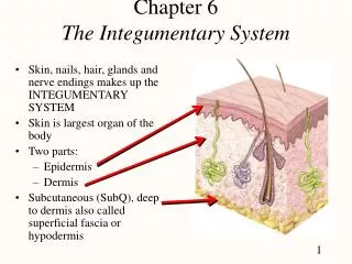
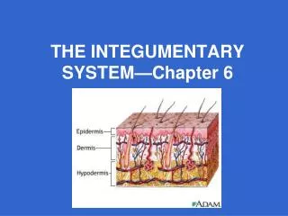
2. 2 Integumentary System = Skin + hair + nails + various glands, muscles and nerves.
Guards physical & biochemical integrity
maintains constant body temperature
provides sensory information
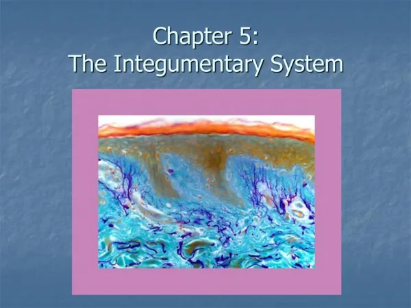
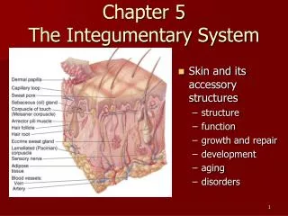
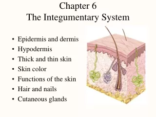
STRUCTURE OF THE SKIN
Largest organ in the body, ~ 2 square meters, 16% of total body weight.
Dematology- Branch of medicine for diagnosing skin disorders.
Skin: 2 main parts
Epidermis
Dermis
3. 3 Epidermis-
Keratinized stratified squamous epithelium
Contains
Keratinocytes- produce keratin, 90% of the cells. Waterproof.
Melanocytes- produce the pigment melanin, 8% of cells. Transfer melanin to keratinocytes where they shield the nuclear DNA from UV rays.
Langerhan cells- participate in the immune response. Easily damaged by UV light.
Merkel cells- least numerous, in the deepest part of the epidermis. Function in the sensation of touch.
Epidermis has 4 layers in most parts of the body except where friction is greatest; (fingertips, palms, soles) there it has 5 layers.
Stratum Basale- deepest layer
4. 4 Composed of a single row of cuboidal or columnar keratinocytes. Some of which are stem cells producing new keratinocytes.
Cytoskeleton of cells in the SB include filaments of keratin which attach to other cells in the stratum spinosum or the basement membrane holding the epidermis to the dermis. (desmosomes, hemidesmosomes)
This is the area that has a role in the formation of new cells.
Stratum spinosum- superficial to the stratum basale
8- 10 layers of keratinocytes fit closely together.
5. 5 Some cells in this layer retain the ability to undergo cell division.
Bundles of intermediate filaments insert into desmosomes, which tightly join the cells to one another, provides both strength and flexibility to the skin.
Projections of both Langerhan cells and melanocytes appear in this stratum.
Stratum Granulosum- the middle of the epidermis.
3-5 layers of flattened keratinocytes undergoing apoptosis. (normal cell death)
We see the presence of darkly stained granules of protein called keratohyalin.
6. 6 Membrane enclosed lamellar granules which release a lipid secretion that fills the spaces between the cells in this layer. Actts as a water repellent and sealant that slows the loss of body fluids and the entry of foreign materials.
The nuclei at this level are dying, the cells can no longer metabolize.
Transition between metabolically active cells and dead cells is made here.
Stratum Lucidum- Only present in the fingertips, palms and soles.
3-5 layers of clear, flat, dead keratinocytes with densely packed intermediate filaments.
7. 7 Stratum Corneum-
25-30 layers of dead, flat keratinocytes
Cells contain mostly dense packed intermediate filaments and keraohyalin.
Between the cells are lipids, from lamellar granules to aid with water repellent nature.
Cells are continually shed and replaced.
Constant friction at this area stimulates the formation of calluses
8. 8 Growth of the epidermis
As cells form in the SB they are pushed to the surface by a process called Keratinization.
As they undergo apoptosis they are replaced by new cells.
The whole process takes ~4 weeks.
Psoriasis- cells are shed prematurely. Immature cells make abnormal keratin and form flaky silver scales on the skin surface. Treatment is to supress cell division.
9. 9 DERMIS- Deeper part of the skin.
Composed mainly of CT containing collagen &elastin fibers
blood vessels, nerves, glands and hair follicles are embedded in dermal tissue.
Has a superficial papillary region and a deeper reticular region.
Papillary region- 1/5 thickness of total layer. Contains CT with fine elastic fibers and fingerlike projections called dermal papillae.
Also found here are meissner corpulses ( nerve endings sensitive to touch) and free nerve endings capable of relaying signals of warmth, cool, pain, tickling, itching.
10. 10 Reticular region_ Dense irregular CT with bundles of collagen and some elastin fibers.
Spaces occupied by adipose cells, hair follicles, nerves, sebaceous glands, and sweat glands.
Strength, extensibility and elasticity. IE. Preganancy, obesity. Stretch marks = tears in the dermis.
Epidermal ridges- series of ridges and grooves on the palms, fingertips, soles and toes.
Increase grip by friction
Small glands form fingerprints, unique to each individual.
11. 11 Basis of Skin color
Melanin, Carotene and hemoglobin- 3 pigments that give skin its color.
The amount of melanin causes skin color to vary from pale yellow to black.
Melanocytes- most evident in mucous membranes, penis, nipples, face and limbs.
Color is based on the amount of pigment the melanocytes produce and disperse to the keratinocytes.
Accumulates in patches called freckles & liver spots.
Melanocytes synthesize melanin from tyrosine with the help of the enzyme tyrosinase. Occurs in the organelle called a melanosome.
UV increases enzymatic activity
12. 12 Carotene- Yellow orange pigment, precursor of vitamin A.
When little melanin or carotene are present the skin of white people appears red due to hemoglobin.
Albinism- Inherited. Unable to synthesize tyrosinase.
Vitiligo- Partial or complete loss of melanocytes. Irregular white patches.
Cyanotic, jaundice, Erythema.
13. 13 Accessory structures of the skin
Hair- protection
Nails- protection
Sweat glands- help regulate temperature.
Hair Anatomy-
Columns of dead, keratinized cells bonded together by extracellular proteins.
Shaft- superficial part of the hair
Root- portion of hair deep to the shaft. Penetrates into the dermis, sometimes into the subcutaneous layer.
Shaft & root consist of 3 concentric layers.
Inner Medula- pigment granules & air spaces
Middle cortex- pigment granules in dark hair, air in gray hair.
Cuticle- outermost layer, flat keratinized cells single layer.
14. 14 The base of hair contains a bulb
Houses a structure called the papilla
Contains CT and many blood vessels that nourish the hair follicle.
Also contains a germinal layer of cells called the matrix, responsible for the growth of new hair when old hair is shed.
Arrector Pilli- smooth muscle bundle associated with hair. Extends from the superficial dermis to the side of the follicle.
Causes �goose bumps� secondary to an autonomic reaction.
Hair growth- Scalp hair grows for ~ 2-6 yrs and rests 3 months. At any time ~85% of scalp hair is growing.
Hair color- Depends on amount & type of melanin present. Gray hair is a lack of tyrosinase.
Function of hair- Protection, sensation of light touch.
15. 15 SKIN GLANDS
Sebaceous (oil)
Sudoriferous (sweat)
Ceruminous
Mammary
Sebaceous- oil glands, simple branched glands.
Connected to hair follicles
Secreting portion is in the dermis and opens into the hair follicle.
Secrete an oily substance called sebum, a mixture of fats, cholesterol, proteins, inorganic salts.
Coats the surface of hair and also helps prevent excessive evaporation of water from the skin. Keeps skin soft & pliable. Inhibits bacteria growth. Ie. Acne
16. 16 Sudoriferous glands- Sweat glands.
Release secretions by exocytosis onto the skin through pores or into hair follicles.
2 main types
Eccrine- Most common, tubular, distributed throughout the skin. Most numerous in the forehead, palms, soles
Secretory portion located deep in the dermis.
Consists of water, ions, urea, uric acid, ammonia, glucose & lactic acid.
Main function is to regulate body temperature through evaporation.
Apocrine- found in the skin of the axilla, groin, nipples, breasts, male bearded regions.
Actually mesocrine glands.
Do not begin to function until puberty. Stimulated during stress
17. 17 Ceruminous- modified sweat glands in the external ear (earwax) Function protection of ear as a sticky barrier.
Nails.- Tightly packed, hard, keratinized epidermal cells.
Nail body- visible portion
Nail root- buried in the fold of the skin.
Nail matrix- epithelium deep to the nail root, cell producing by mitosis.
Types of skin
thin (hairy)- covers all parts of body except palms, soles, digits. Has fewer sweat glands than thick skin.
18. 18 Thick skin-
lacks hair follicles, arrector pilli muscles ans sebaceous glands.
Has densely clustered sensory receptors.
Functions of skin
Thermoregulation- Homeostasis
protection
Cutaneous sensations- touch, pressure, vibration, warmth, coolness.
Excretion & absorption- ~400 ml water evaporates. Absorbs fat soluable vitamins A,D,E,K.
Synthesis of vitamin D requires activation of a precursor molecule in the skin by UV rays.
19. 19 Skin Wound healing-
Epidermal- abrasions, minor burns.
Deep wound healing- extends into the dermis.
Inflamatory phase- blood clot forms, vascular & cellular response
Migratory phase- clot becomes a scab. Cells migrate beneath the scab to bridge the wound.
Granulation tissue- fills the wound at this point
Proliferative phase- Extensive growth of epithelial cells under the scab. Continued growth of blood vessels.
Maturation phase- Scab falls off, epidermis restored to normal thickness. Blood vessels restored to normal.
Hypertrophic scar vs. keloid scar
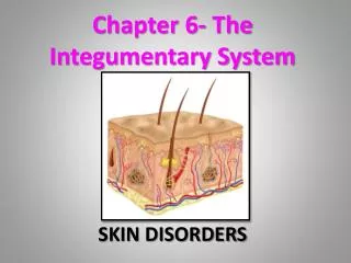
20. 20 Skin Cancer-
Basal cell carcinoma- ~78% of all skin cancer. Tumor arises in stratum basale.
Squamous cell carcinoma- ~20%
Malingnant melanoma- ~2% Arises from melanocytes. Most life threatening in young women Risk of 1 in 75.
The key to treatment is early detection
Burns
1st degree- only epidermis
2nd degree- possibly dermis is involved.
3rd degree- full thickness, epidermis, dermis and some underlying structures.
Rule of nines- used to estimate surface area of a burn.
21. 21 Finally done!!