Download
1 / 22
230 likes | 402 Vues
Traveling Through Proteomes Using 3D-EM and AFM. Nanoanalysis, July 10, 2006, ETHZ Andreas Engel Maurice E. Müller Institiute, Biozentrum University of Basel, Switzerland. Electron Tomography. 2D-projections => 3D-reconstruction. Principle of electron tomography.

E N D
Traveling Through Proteomes Using 3D-EM and AFM Nanoanalysis, July 10, 2006, ETHZ Andreas Engel Maurice E. Müller Institiute, Biozentrum University of Basel, Switzerland
2D-projections => 3D-reconstruction Principle of electron tomography 3D-object => set of 2D-projections W. Baumeister, R. Grimm, J. Walz: Trends Cell Biol 9 (1999) 81-85
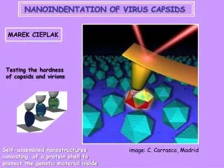
Apical part of a sporozoite cell membrane polar rings microtubules ER rhoptries micronemes dense granules
Nanoanalytics of Soluble Complexes:Scanning Transmission Electron Microscopy (STEM)
Scanning Transmission EM 200 Å Philippe Ringler
BF DF PM STEM Hardware Single electron counting Beam current Acceleration voltage 100 kV Pressure: < 10-10 Torr
14 nm TMV Analysis
Actin Collaboration with Ueli Aebi, M.E. Müller Insitute
From mass to shape Mueller et al, J Mol Biol 99 STEM
Nanoanalytics of Membrane Complexes:Atomic Force Microscopy (AFM)
Membrane Proteins exist in the Bilayer Bert de Groot & Helmut Grubmüller
Cytosolic Surface of Bacteriorhodopsin Dimitrios Fotiadis, unpublished
CS of Bacteriorhodopsin: Force-induced Conformational Changes 10 nm (A), 4 nm(B, C and D) Müller et al. (1995), J. Mol. Biol. & Fotiadis et al. (2002), Micron
The Surface Dynamics of Bacteriorhodopsin Similarity ranked images are assembled into a movie Low force High force Scheuring et al., European Biophysics Journal
Bacteriorhodopsin: Surface Energy Landscape pd(r) peak position probability of domain d Low force High force Fd = -kTlnpd(r) Scheuring et al. Eur Biophys J 2002 6 kT Low force High force
Unzipping Bacteriorhodopsin Oesterhelt et al. Science 2000
Conclusions • Electron and atomic force microscopies offer great tools for cellular nanoanalytics • Electron tomography provides entire picture of a cell • STEM makes the link between mass and shape • AFM is an ideal tool for assessing structure & dynamics of membrane proteins
AFM Daniel Müller* Simon Scheuring* Dimitrios Fotiadis Patrick Frederix Acknowledgments Protein expression Nora Eifler Myriam Duckely Paul Werten 2D crystallization Hervé Rémigy Thomas Kaufmann Thomas Walz* Peter Agre Wolfgang Baumeister Yoshi Fujiyoshi Helmut Grubmüller Bert deGroot Kris Palczewski STEM Shirley Müller Philippe Ringler Francoise Erne-Brand