Download
1 / 26
260 likes | 266 Vues
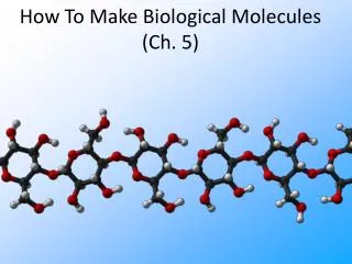
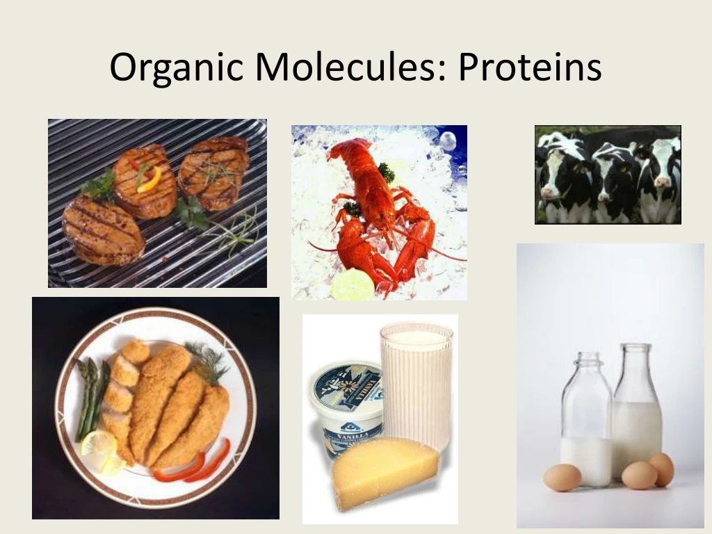
Organic Molecules: Proteins. Proteins. Most structurally & functionally diverse group Function: involved in almost everything enzymes (pepsin, DNA polymerase) structure (keratin, collagen) carriers & transport (hemoglobin, aquaporin) cell communication

E N D
Proteins • Most structurally & functionally diverse group • Function: involved in almost everything • enzymes (pepsin, DNA polymerase) • structure (keratin, collagen) • carriers & transport (hemoglobin, aquaporin) • cell communication • signals(insulin & other hormones) • receptors • defense (antibodies) • movement (actin & myosin) • storage (bean seed proteins)
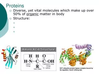
H2O Proteins • Structure • monomer =amino acids • 20 different amino acids • polymer =polypeptide • protein can be one or more polypeptide chains folded & bonded together • large & complex molecules • complex 3-D shape hemoglobin growthhormones Rubisco
Amino acids • Structure • central carbon • amino group • carboxyl group (acid) • R group (side chain) • variable group • different for each amino acid • confers unique chemical properties to each amino acid - like 20 different letters of an alphabet - can make many words (proteins) Oh, I get it! amino = NH2 acid = COOH
Effect of different R groups: Polar amino acids • polar or charged & hydrophilic H+ acceptors H+ donors Why are these polar & hydrophillic?
Effect of different R groups: Nonpolar amino acids • nonpolar & hydrophobic Why are these nonpolar & hydrophobic?
Sulfur containing amino acids • Formdisulfide bridges S-S • covalent cross links betweens sulfhydryls • stabilizes 3-D structure H-S – S-H
dehydration synthesis peptidebond H2O Building proteins • Peptide bonds • covalent bond between NH2 (amine) of one amino acid & COOH (carboxyl) of another • C–N bond AP Biology
Building proteins • Polypeptide chains have direction • N-terminus = NH2 end • C-terminus = COOH end • repeated sequence (N-C-C) is the polypeptide backbone • can only grow in one direction http://www2.nl.edu/jste/proteins.htm
hemoglobin collagen Protein structure & function • Function depends on structure • 3-D structure • twisted, folded, coiled into unique shape pepsin
Protein Structure • Protein types include globular proteins which are usually enzymes and Fibrous proteins which usually serve for structure (eg. Hair) • Proteins Exhibit 4 levels of structure.
Primary (1°) structure • Order of amino acids in chain • amino acid sequence determined by gene (DNA)l; dictates all further levels of protein structure • slight change in amino acid sequence can affect protein’s structure & its function • even just one amino acid change can make all the difference!
Sickle cell anemia Just 1out of 146amino acids! I’mhydrophilic! But I’mhydrophobic!
Normal hemoglobin Sickle-cell hemoglobin Primary structure Primary structure . . . . . . Exposed hydrophobic region Val His Leu Thr Pro Glul Glu Val His Leu Pro Glu Thr Val 5 6 7 3 4 5 6 7 1 2 1 2 3 4 Secondaryand tertiarystructures Secondaryand tertiarystructures subunit subunit Quaternary structure Hemoglobin A Quaternary structure Hemoglobin S Molecules interact with one another tocrystallize into a fiber, capacity to carry oxygen is greatly reduced. Function Molecules donot associatewith oneanother, eachcarries oxygen. Function 10 m 10 m Normal cells arefull of individualhemoglobinmolecules, eachcarrying oxygen Red bloodcell shape Red bloodcell shape Figure 5.21 Fibers of abnormalhemoglobin deform cell into sickle shape.
Secondary (2°) structure • “Local folding” • folding along short sections of polypeptide • interactions between adjacent amino acids • H bond: weak bonds between R groups • forms sections of 3-D structure • -helix • -pleated sheet
Tertiary (3°) structure • “Whole molecule folding” • interactions between distant amino acids • hydrophobic interactions • cytoplasm is water-based • nonpolar amino acids cluster away from water • Covalent, H bonds & ionic bonds • disulfide bridges • covalent bonds between sulfurs in sulfhydryls (S–H) • anchors 3-D shape
Quaternary (4°) structure • More than one polypeptide chain bonded together • only then does polypeptide become functional protein • pH, changes or heat can disrupth bonds perm. denaturing the protein hemoglobin collagen = skin & tendons
Protein structure (review) R groups hydrophobic interactions disulfide bridges (H & ionic bonds) 3° multiple polypeptides hydrophobic interactions 1° amino acid sequence peptide bonds 4° 2° determinedby DNA R groups H bonds
Protein denaturation • Unfolding a protein • conditions that disrupt H bonds, ionic bonds, disulfide bridges • temperature • pH • salinity • alter 2° & 3° structure • alter 3-D shape • destroys functionality • some proteins can return to their functional shape after denaturation, many cannot http://highered.mcgraw-hill.com/sites/0072943696/student_view0/chapter2/animation__protein_denaturation.html
Correctlyfoldedprotein Polypeptide Cap Hollowcylinder The cap attaches, causing the cylinder to change shape insuch a way that it creates a hydrophilic environment for the folding of the polypeptide. The cap comesoff, and the properlyfolded protein is released. Steps of ChaperoninAction: An unfolded poly- peptide enters the cylinder from one end. Chaperonin(fully assembled) 2 3 1 Figure 5.23 • Chaperonins • Are protein molecules that assist in the proper folding of other proteins
0 Review Questions
0 A. What happens when a protein denatures? * • It loses its primary structure. • It loses its secondary and tertiary structure. • It becomes irreversibly insoluble and precipitates. • It hydrolyzes into component amino acids. • Its hydrogen bonds, ionic bonds, and peptide bonds are disrupted.
0 B. The R group or side chain of the amino acid serine is –CH2 –OH. The R group or side chain of the amino acid alanine is –CH3. Where would you expect to find these amino acids in globular protein in aqueous solution? • Serine would be in the interior, and alanine would be on the exterior of the globular protein. • Alanine would be in the interior, and serine would be on the exterior of the globular protein. • Both serine and alanine would be in the interior of the globular protein. • Both serine and alanine would be on the exterior of the globular protein. • Both serine and alanine would be in the interior and on the exterior of the globular protein.