Download
1 / 58
600 likes | 878 Vues
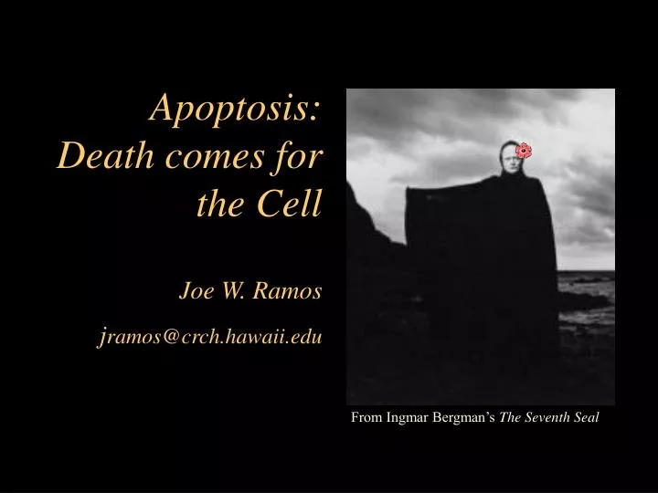
Apoptosis: Death comes for the Cell Joe W. Ramos j ramos@crch.hawaii.edu. From Ingmar Bergman’s The Seventh Seal. Mutations in proteins that regulate cell proliferation, survival and death can contribute to oncogenesis. From Okada and Mak, Nat. Rev. Cancer 4:592-603.

E N D
Apoptosis:Death comes for the CellJoe W. Ramosjramos@crch.hawaii.edu From Ingmar Bergman’s The Seventh Seal
Mutations in proteins that regulate cell proliferation, survival and death can contribute to oncogenesis
Apoptosis: Programmed Cell Death • A term used to describe the morphological changes associated with programmed cell death. • The term was originally used by Wyllie and his colleagues and is from the Greek meaning “dropping away” as the leaves from a tree.
Apoptosis • Activecell death • Requires energy and RNA and protein synthesis • Characteristic morphological features • DNA cleaved, chromatin condenses • Cells shrink • Formation of apoptotic body • Cleared by phagocytosis • No inflammation=no tissue damage
Necrosis • Passive cell death • Cells swell up • Membrane breaks down and cellular contents leak out • Nucleus disintegrates • Cell ghosts • Inflammatory=tissue damage
The function of Cell death • Multicellular development • involved in deletion of entire structures, • sculpting of tissues, • and regulates the neuron number • The immune response • The body’s defense against cancer
Death and the mouse’s paw Dark Green fluorescence indicates apoptotic cells. Fig 18-18
Apoptosis regulates nerve cell targeting Fig 18-20
Detection of apoptotic cells • Microscopy • Cells have classic features (eg. small darkly stained nuclei) • Detection of free 3’ ends of DNA by TUNEL assay (terminal deoxytransferase-mediated dUTP-biotin nick end labeling) • Gel electrophoresis • Detect DNA ladder of 180 bp intervals caused by internucleosomal DNA cleavage • Flow cytometry • Measure externalization of phosphatidylserine (PS) with fluorescently labeled Annexin-V • Measure DNA fragmentation with propidium iodide fluorescence
Analysis of DNA content with a flow cytometer Recall the fluorescence intensity of the DNA dye (amount of DNA) is measured for each cell.
Triggers of apoptosis • Programmed cell death in which many more cells are produced than survive (e.g. development of lymphocytes) • Toxic stimuli (viruses, chemicals, ionizing radiation) • Extracellular signals (Fas, p75 NGF-R, TNF) • DNA damage (p53)
C. elegans has played a key role in our understanding of Apoptosis 1090 total cells 131 die Ced-3=no death ced-1 mutant (No engulfment) Ced-4=no death Ced-9=all die ced-1/ced-3 (No cells die) H.R Horvitz and colleagues responsible for much of this work, 2002 Nobel Prize in Medicine with Sulston and Brenner.
C. elegans apoptosis CED-9=Blocks apoptosis CED-4=linker molecule forms activating complex with CED-3 CED-3=Protease that executes cell by chewing up proteins EGL-1=Proapoptotic by blocking CED-9 function
Three classes of proteins function in the apoptotic pathway-conserved in vertebrates Mammalian Bcl-2 can substitute for Ced-9 in c. elegans
Death’s Methods: A protease cascade These proteases are called caspases Fig 18-22
Caspases • Caspases are Cysteine directed proteases that cleave after ASPartate residues • Ced-3 is the C. elegans homologue • At least 14 family members • Synthesized as proenzymes with low levels of caspase activity (~1-2 % of active form) • Activated upon after aggregation or cleavage to mature form • Caspases –8 and –9 are “initiator” caspases • Caspases –3 is the “effector” caspase • Caspase activation requires a stimulus • They proteolyze cellular proteins to carry out cell death program
Two Pathways that Initiate Apoptosis • Intrinsic/ Mitochondrial Apoptosis • Regulated by Mitochondria • Cytochrome c release • Extrinsic/ Death Receptor Apoptosis • Activated by ligation of Death Receptors • Fas, TNF alpha • These pathways intersect at the effector caspases
Intrinsic/Mitochondrial Pathway CARD domain
Intrinsic Pathway: Apaf-1 Induced Apoptosis CARD domains
Smac/Diablo and IAPs Smac=Second mitochondrial activator of caspases IAP=Inhibitor of Apoptosis Proteins
Bcl-2 family members • A very large family with 19 members identified • Bcl-2 (homologous to ced-9) is prototype • All have the BH3 domain (Bcl-2 Homology) • BH-3 is the pro-apoptotic domain exposed on activation • Act as dimers=either hetero or homodimers • Pro-apoptotic dimers (Bax) increase mitochondrial permeability • Anti-apoptotic members (Bcl-2, Bcl-XL) form dimers with pro-apoptotic members to inactivate them
The Bcl-2 Family BH domains=protein-protein interaction domains
Some trophic factors prevent apoptosis by inducing inactivation of a pro-apoptotic regulator Figure 23-50
Mitochondrial permeability PT=Permeability transition, bursts outer membrane
Cell, Vol 111, 331-342, 1 November 2002Bid, Bax, and Lipids Cooperate to Form Supramolecular Openings in the Outer Mitochondrial Membrane Tomomi Kuwana 1, Mason R. Mackey 2, Guy Perkins 2, Mark H. Ellisman 2, Martin Latterich 3, Roger Schneiter 4, Douglas R. Green 1, and Donald D. Newmeyer 1
Bax+ BH3 Peptide (Direct Activation) Bax + N/C-Bid + Bcl-xL+ BH3 Peptide (De-repression) * * * Bid and Bad have distinct functions to activate apoptosis Liposome Assay (cardiolipin+Bax+Bid) tBid Directly activates Bax pore formation Bad indirectly activates Bax pore formation (Binds Bcl-xL→ releasing Bax) N/C-Bid=recombinant activated Bid BH3 Peptide Kuwana et al., Molecular Cell, 17, 525-535, 2005
Death Receptors and Ligands CD95=Fas
TNF receptor family Cysteine-Rich Domains (CRD) Death Domains (DD) Bind DDs of other proteins (e.g. FADD)… …Recruiting them to the plasma membrane.
Fas-FasL Apoptosis • In response to antigenic stimulation, peripheral T cells expand • The antigen specific T cells generated must be eliminated (except for the memory cells) • Upon repeated antigenic stimulation via the T Cell receptor: T cells upregulate Fas and FasL • Eliminate neighboring T Cells expressing Fas
Fas Induced Apoptosis The Formation of the Death Initiating Signal Complex (DISC)
Adaptor Proteins contain conserved protein interaction domains = inhibits apoptosis -CARD domain of Apaf-1 binds CARD domain of procaspase-9. -DED domain of FADD binds DED domain of procaspase-8. -DED domains of FLIP can bind to the DED domain of FADD and block procaspase-8 recruitment.
Proteolytic targets of effector caspases • Cytoskeletal regulatory proteins • Actin • Nuclear Lamins • Poly(ADP-ribose) polymerase (PARP) • PARP activity depletes ATP, thus cleavage of PARP may maintain store of ATP to drive apoptosis • DNA-fragmentation factor (DFF)
Two roads to activate apoptosis Extrinsic Intrinsic
TNF receptors also signal to NFkB Ubiquitylation is a common signal transduction mechanism (see regulation of cyclins for example) IKKK=IkB Kinase kinase NFkB activates transcription of several anti-apoptotic proteins including IAPs and Bcl-2.