Download

1 / 27
270 likes | 392 Vues
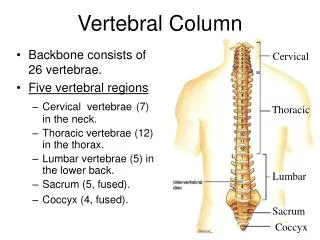
Histiocytosis and vertebral involvement. a 6 year old boy child CC : Back pain since 6 MO ago( 3/91) Lumbar X Ray: Nl 3/91 Evaluation : 19/6/91 WBC: 9.8 ( PMN:%74 L:% 24 E :%2 ) HB: 11.7 Plt : 302 ESR: 30 CRP: 25.9 ( 0-6)

E N D
a 6 year old boy child • CC: Back pain since 6 MO ago( 3/91) • Lumbar X Ray: Nl 3/91 • Evaluation : 19/6/91 • WBC: 9.8 ( PMN:%74 L:%24 E:%2 ) • HB: 11.7 Plt: 302 • ESR: 30 CRP: 25.9 ( 0-6) • Wright : Negcoombswright : Neg • CXR:NL History
AbdSono: NL • Bone Scan: active bone lesion of the L4 spine. 25/6/91 • Spiral CT scan :Destructiv Lesion on antero – lateral aspect of L4 vertebral body with mild prevertebral soft tissue component. Spinal canal is intact. 26/6/91 • Lumbo-sacral MRI : HypersignalT2W hyposignal T1W L4 body lesion, needs MRI with IV gadolinim for ruling out infiltrative lesion , posterior or postero-lateral disc :No displacement History
Needle Biopsy- guide of CT Scan Report : LCH 4/7/91 • IHC : Difffuse positive reaction for S- 100 protein and CD1A,Vimentin and CD68 • Review of HP: LCH • Treatment: Brace • Follow up : ClinicalyStable, No back pain • Bone survey: ( after 2 Month): NL 29/8/91 History
WBC: 6.96 PMN= %63.3 L= %29.7 M:%5 E: %1.7 B :% 0.3 Hb= 12 MCV: 80 MCH: 28 Plate: 302 Retic: 0.3% D& I coombsTest: Neg ESR:8(up to 10) CRP: 5( up to 5) U/A: SG:1020 BS: 97 BUN :37 Creat: 0.46 Calcium: 9 P: 5.1 ALK:417 SGOt: 31 SGPT: 15 BILT: 0.6 Bil D:15 T-protein: 6.7 S- Alb: 4.4 PT= 13.8 INR:1.09 PTT: 30 HBSAg: neg HBS AB: 10.6 HIV: NegHCVAb:Neg TSH: 3.47 T4: 8.8 Ferritin: 32.3 HbElecthrophorasis: Nl 1/9/91 Next MRI: NEXT month( 10/91) History
Up to 80% of LCH lesions in children are of EG type. • The incidence of spinal involvement : 6.5% to 25% • 80% of cases : children <10 years of age • usual appearance of the spinal LCH is vertebral body osteolytic lesions that leads to “vertebra plana” • LCH may be more aggressive, which may make the lesions look like a malignancy, which appear as bubbly and lytic lesions, and expand posterior elements with a soft tissue mass. Histiocytosis
Reports on para vertebral soft tissue mass in LCH of the spine are rare in the literature • Since the introduction of (MRI), soft tissue extension of vertebral body LCH become a well known entity • the incidence of soft tissue extension is unknown because reports are either case reports or reviews • Patients easily Misdiagnosed with malignant tumours, more aggressive tumours, lymphom or tuberculosis . • vertebra plana with soft tissue mass can be also seen in variety of spine tumours, including Ewing’s sarcoma, lymphoma, aneurysmal bone cyst, leukemia, malignant solid tumours. Histiocytosis
The treatment of LCH of the spine with soft tissue extension, is still controversial. • treatments of LCH of the spine: • prolonged bed rest, • immobilisation with cast and brace, • Chemotherapy, • Radiation therapy and surgery • neurological deficit • Most researchers suggested that the neural elements should be decompressed and then fused • Some investigators advocated immobilisation and radiation • A few recommended hormone or chemotherapy Histiocytosis
1980 -2003 , 8children with LCH <18 years of age, ,vertebral involvement • Most common presenting :back or neck pain. • Thoracic vertebrae most commonly affected followed equally by cervical and lumbar spines. • Most children underwent a complete diagnostic work-up. • Biopsies were attempted in all cases with 6 positive results. • Treatment depend on the severity of the presenting complaint; • None of the tumours was completely resected. Follow-up averaged 3.4 years Only 1 child has had a recurrence. Treatment and outcome of vertebral Langerhans cell histiocytosisat the Children’s Hospital of Eastern Ontario Christopher W. J can chir, Vol. 48, No 3, juin 2005
Case 3. There is 50% canal compromise at the L4 vertebra. The lateral radiograph (A) demonstrates the classic vertebra plana of the anterior vertebral bodof L4. The bone scan (B) shows uptake of contrast at L4. CT (C) and MRI (D,E) images further demonstrate the degree of canal compromise at this leve
FIG. 2. Case 8. Sagittal (A) and transverse (B) MR images demonstrate the solitary lesion of Langerhans cell histiocytosis involving the spinousprocess of T12 as well as the paraspinalsoft tissuse.
FIG. 3. Case 8. Histologic sections show an atypical histiocytic infiltrate (A; hematoxylin-phloxine-saffron stain, original magnification ×25), eosinophils in the background(B; Giemsa stain, original magnification ×25), positive CD1a staining (C;original magnification ×25) and Birbeck granules on electron microscopy (D; originalmagnification ×55 000).
DIAGNOSIS • Although some maintain that radiographically typical lesions do not require biopsy and can safely be followed with serial radiography, Most advocate a tissue diagnosis • Any child with suspected solitary LCH of the vertebra undergo a full diagnostic investigation consisting of a skeletal survey / Abdominal ultrasonography • Although bone scanning is not sensitive for LCH, it is also recommended because it is important to rule out other diagnoses. • CT : vertebral lesion • MRI : Neural involvement. • A biopsy is highly recommended for a diagnosis Summary and conclusions
Treatment of LCH in the spine • immobilization and observation: in the absence of multisystem disease or instability • Surgery: instability or neurologic Deficit. • Chemotherapy : systemic involvement. • Radiotherapy: There is no role for routine radiotherapy. unless the disease continues to progress. • Follow-up should be a minimum of 3 years or until symptoms or the lesion has resolved.
A rare case of C-6 vertebral involvement in a 12-year-old boy with histiocytosis X. The patient presented with limitation in movements of neck and upper extremities. Surgery was performed via an anterior cervical approach along with stabilization using a fibula strut graft and plate fixation. HP Report : LCH The patient went on to make a full recovery with complete resolution of his motor weakness. Rare C-6 vertebral involvement in a child with histiocytosis X: Case report Folia Neuropathol 2007; 45 (2): 93-97
Rare C-6 vertebral involvement in a child with histiocytosis X: Case report Folia Neuropathol 2007; 45 (2): 93-97
Discussion • Gandolfi :spinal cord compression duo to vertebral body of T-7, which was surgically corrected. • Boriani et al 35 cases of vertebral histiocytosis with a total of 52 vertebrae affected. They noted that the diagnosis was based on radiology only in typical cases; and in cases of malignant neoplastic lesions, the diagnosis might be missed. These authors proposed orthotic stabilization and/or surgical treatment based on the extent of the lesion and the potential for further progression. • Kayser et al:6 children LCH who underwent operative treatment. These authors also noted that in cases of vertebral Langerhans cell histiocytosis, differentiating these lesions from spinal osteomyelitis may be difficult. Rare C-6 vertebral involvement in a child with histiocytosis X: Case report Folia Neuropathol 2007; 45 (2): 93-97
Abstract : Incidence of obvious soft tissue extension from LCH of the spine in children and to evaluate the effects of chemotherapy for those patients. 18 patients LCH /9 with obvious paravertebral soft tissue extension 8 patients / neurological symptoms. 3 typical “vertebra plana” All received chemotherapy and one had surgical treatment. The mean follow-up time was 30.3 months. Soft tissue extension disappeared completely in all patients. No clinical evidence of disease was observed at the most recent follow-up. Chemotherapy is safe and effective, and surgical decompression was probably not necessary for most patients Langerhans’ cell histiocytosis of the spine in children with soft tissue extension and chemotherapInternational Orthopaedics (SICOT) (2009) 33:731–736 Xin-Sheng Peng
Fig. 1 Atypical radiographical presentation of the vertebral involvement of Langerhans’ cell histiocytosis (LCH) of the spine in children with soft tissue mass. One lateral erosion with soft tissue mass of T7case 7
Fig. 2 LCH of T7. Axial computed tomography (CT) image demonstrating circumferential soft tissue extension from the vertebrabody(case 6)
Chemotherapy agents and regimens used to treat LCH of the spine with obvious soft tissue extension in children Vincristine0.5–1 mg/m2 IV, per week (3 months) IV, per two weeks (3 months) IV, per four weeks (3 months) Methotrexate 5–10 mg/m2 IV, per week (3 months) IV, per two weeks (3 months) IV, per four weeks (3 months) Prednisone 5 mg/m2 Oral, per day (9 months) 6-Mercaptopurine 10 mg/m2 Oral, per day (6 months) 5 mg/m2 Oral, per day (3 months) Langerhans’ cell histiocytosis of the spine in childrenwith soft tissue extension and chemotherapy International Orthopaedics (SICOT) (2009) 33:731–736 Xin-ShengPeng
Fig. 5 LCH of T4. MRI views (T1-weighted with gadoliniumenhancedimage) shows the prominent soft tissue mass intruding intothe spinal canal, which disappeared completely one month afterchemotherapy (case 8)
Conclusion: • chemotherapy is a safe and effective method to treat patients with LCH of the spine in children with soft tissue Extension. • In this study :chemotherapy was able to rapidly reduce the intraspinal soft tissue mass and to relieve the local pain and radicular pain, even with spinal cord and nerve root compression. • 8/9 patients were cured only by chemotherapy • there was no evidence that chemotherapy is a good option in the management of LCH of the spine in children with soft tissue extension, especially the patients with neurological deficits. • surgical decompression is probably not necessary for most patients • Only with spinal instability or neurological deficits caused by kyphosis is surgical treatment recommended Langerhans’ cell histiocytosis of the spine in childrenwith soft tissue extension and chemotherapy International Orthopaedics (SICOT) (2009) 33:731–736 Xin-ShengPeng
Spine (Phila Pa 1976). 1998 Jun 15;23(12):1351-4. Vertebral remodeling in eosinophilicgranuloma of the spine. A long-term follow-up. Raab P, Hohmann F, Kühl J, Krauspe R. Source Department of Orthopaedic Surgery, University of Würzburg, Germany. Abstract STUDY DESIGN: In this study, 14 conservatively treated patients were reviewed who had eosinophilicgranuloma of the spine, which had been diagnosed on the basis of histologic study of the vertebral lesion or of specimens from other sites in patients with multiple involvement. The remodeling of the vertebral body was studied in an average follow-up of 5.6 years. mandatory for an accurate diagnosis. Abstract 14 patients with eosinophilicgranuloma of the spine, on the basis of histologic diagnosis who was treated conservatively The remodeling of the vertebral body was studied in an average follow-up of 5.6 years vertebral height was calculated by measuring the ventral distance between the superior and inferior margins of the vertebral body cervical spine in 2 patients and in thoracic and lumbar vertebrae in 7 patients each. Vertebral remodeling in eosinophilic granuloma of the spine. A.long-term follow-up.Department of Orthopaedic Surgery, University of Würzburg, Germany1998 Jun 15;23(12):1351-4.
Conclusion: conservative orthopedic treatment with immobilization in a brace is sufficient to allow for optimal vertebral remodeling. Partial to nearly complete reconstitution of vertebral height was seen in all cases. Thus, operative treatment with curettage and bone grafting is not necessary. In instances with neurologic impairment surgical decompression and short fusion of the spine is necessary. Nevertheless, biopsy are mandatory for an accurate diagnosis .Vertebral remodeling in eosinophilicgranuloma of the spine. A long-term follow-up.
Abstract 26 children with biopsy-proven LCH involving the spine were treated between 1970 and 2003. Vertebral body collapse was measured on radiographs and classified as grade I (0% to 50% collapse) or grade II (51% to 100% collapse) and subclassified as A (symmetric collapse) or B (asymmetric collapse). Lesions of the posterior elements of the spine were classified as grade III. Twenty-three children were followed for two years or more (mean, 9.4 years), Langerhans cell histiocytosis of the spine in children. Long-term follow-up.Children'sHospital of Philadelphia, Philadelphia, USAJ Bone Joint Surg Am. 2004 Aug;86-A(8):1740-50.
RESULTS: (62%) children were found to have multifocal skeletal disease.. The extent of the initial collapse seen radiographically was grade IA for twenty vertebrae, IB for three, IIA for ten, IIB for nine, and III for two. Grade-I lesions were more likely to be associated with symmetric collapse than were grade-II lesions. Spinal deformity developed in four children, and two later required spinal fusion. No relationship was observed between the grade of the initial collapse and the subsequent development of spinal deformity. Despite heterogeneous treatment, all patients were alive and well with resolution of all presenting signs and symptoms and no evidence of active disease at the time of the most recent follow-up. CONCLUSIONS: . The natural history of these lesions in the spine in the absence of systemic disease or spinal deformity is such that aggressive surgical management is usually not indicated; only follow-up is necessary to monitor recovery and spinal balance.