Download
1 / 192
1.96k likes | 2.21k Vues
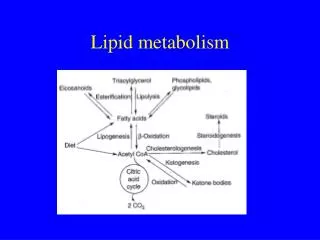
Lipid metabolism. You give me fever Peggy lee . FATTY ACID OXIDATION β -oxidation is the pathway by which the fatty acids from both dietary fat and adipose tissue TAG are oxidized to acetyl- CoA .

E N D
Lipid metabolism You give me fever Peggy lee
FATTY ACID OXIDATION • β-oxidation is the pathway by which the fatty acids from both dietary fat and adipose tissue TAG are oxidized to acetyl-CoA. • The reducing equivalents released during fatty acid oxidation are captured in the form of FADH2 and NADH, which are used to support oxidative phosphorylation • In most circumstances, the acetyl-CoA units generated by β -oxidation will subsequently be oxidized through the TCA cycle, generating additional FADH2 and NADH and ultimately, additional ATP • All cells and tissues except red blood cells and the brain oxidize fatty acids to generate ATP • The β-oxidation pathway is absent in red blood cells because they lack mitochondria • Although neuronal cells in the brain do contain mitochondria, there is only limited transport of fatty acids across the blood-brain barrier
The skeletal and heart muscles in particular have a large capacity for oxidizing fatty acids • Normally, 60 to 90% of the energy required for contraction of the heart is derived from the oxidation of fatty acids • In the fasted state, many cells and tissues depend on β-oxidation of fatty acids to provide the ATP needed to maintain ion gradients and to support biosynthetic processes such as gluconeogenesis • During prolonged fasting or starvation the brain meets its energy needs by oxidizing ketone bodies as well as glucose • The ketone bodies of physiological significance are four-carbon anions produced from acetyl-CoA generated by the β-oxidation of long-chain fatty acids in the liver • Oxidation of ketone bodies by the brain reduces the brain’s dependence on glucose and thus decreases the body’s need to catabolize muscle proteins for gluconeogenesis
The major fatty acids of both dietary TAG and adipose stores in the body contain 16 to 18 carbons and are oxidized through mitochondrial β-oxidation • Essentially the same pathway is utilized for short- (C4-C6) and medium-chain (C8-C12) fatty acids • Related peroxisomal pathways oxidize the less common branched-chain and very long-chain (>C22) fatty acids (VLCFA) • Transport and Activation of Fatty Acids • Since long-chain fatty acids are poorly soluble in aqueous media, they must be transported in the plasma complexed with albumin • When a fatty acid dissociates from albumin it is transferred from the capillary lumen through the capillary endothelium and interstitial space to the cells below • Long-chain fatty acids enter cells both by simple diffusion and by carrier-mediated transport
Intracellularly, fatty acids are bound to cytosolic fatty acid binding proteins (FABP), which deliver the fatty acids to the sites where they are metabolized • Once inside cells, fatty acids must be activated before they can be metabolized • In contrast to glucose, which is activated and trapped within cells as glucose 6-phosphate,fatty acids are converted not to acyl phosphates but to thioesters of coenzyme A • Long-chain fatty acids destined for β-oxidation are activated to their CoA forms primarily on the surface of the outer mitochondrial membrane • The inner mitochondrial membrane is, however, impermeable to long-chain fatty acyl-CoA molecules • Transport of LCFA across the inner mitochondrial membrane is facilitated by a fatty acid transport mechanism called the carnitine shuttle
Carnitine is a quaternary amine that has a hydroxyl group to which a fatty acid can be attached • Since carnitine can be synthesized in the liver and kidney, it is not usually considered an essential dietary nutrient • The activity of the carnitine translocase system is dependent on two enzymes, carnitine palmitoyltransferase I (CPT-I) and carnitine palmitoyltransferase II (CPT-II), both of which catalyze the reversible transfer of long-chain fatty acids between CoA and carnitine • CPT-I is localized to the mitochondrial outer membrane and acts to generate acylcarnitine • Carnitine translocase, which is embedded in the inner mitochondrial membrane transports acylcarnitine into the mitochondria in exchange for free carnitine, which is concurrently exported from the mitochondrial matrix into the cytosol
CPT-II, which is localized to the matrix face of the inner mitochondrial membrane, forms mitochondrial acyl-CoA from CoASH and acylcarnitine • The net effect is the transfer of a long-chain fatty acid from an cytosolic CoASH to a mitochondrial CoASH • Intramitochondrialβ-Oxidation • The term β-oxidation is derived from the fact that the critical chemistry of the four core reactions that comprise the pathway takes place on the third carbon from the carboxyl end: that is, the β-carbon atom • The first of the four reactions of the β-oxidation pathway is irreversible and is catalyzed by acyl-CoA dehydrogenase • Two hydrogen atoms are removed-one each from the αandβ and -generating a carbon-carbon double bond between the αandβ carbons • These hydrogen atoms are transferred to FAD to form FADH2
The Mechanism of The Carnitine Shuttle
Mitochondria contain a family of FAD-linked acyl-CoAdehydrogenases: a long-chain acyl-CoA dehydrogenase, a medium-chain acyl-CoA dehydrogenase and a short-chain acyl-CoA dehydrogenase • The second step in β-oxidation involves hydration of the carbon-carbon double bond between by enoyl-CoAhydratase. The hydroxyl group is introduced onto the β-carbon • In the third step, a second dehydrogenase, NAD +-linked β-hydroxyacyl-CoA dehydrogenase, oxidizes the hydroxyacyl-CoA molecule to generate a β-ketoacyl-CoA and a molecule of NADH • The fourth and final step in β-oxidation involves cleavage of the fatty acid chain with attachment of a second molecule of CoASH to the β-carbon and generation of one molecule of acetyl-CoA • The enzyme that catalyzes this reaction is called β-ketoacyl-CoAthiolase
The net effect of the four steps in β-oxidation is the production of one molecule of acetyl-CoA and one fatty acyl-CoA molecule whose carbon chain is two carbons shorter than the original substrate • The four steps are then repeated, with successive chain shortening by two carbon atoms • The final thiolytic cleavage reaction converts the 4-carbon β -ketobutyryl-CoA (acetoacetyl-CoA) into two molecules of acetyl-CoA • The Energy Yield from β-oxidation • A cycle of β-oxidation releases one molecule each of FADH2, NADH and acetyl-CoA • The four steps are repeated n/2 - 1 times ( n = number of carbons) until the final acetyl-CoA is released • n/2 acetyl-CoA and n/2 - 1 each of FADH2 and NADH are produced by the complete oxidation of a fatty acid with an even number of carbons
Taking the oxidation of palmitate : • Palmitoyl-CoA+ 7 CoA+ 7 FAD+ 7 NAD++7 H2O→ 8 acetyl-CoA+ 7 FADH2+7 NADH+ 7H+ • 8 acetyl-CoA ≈ 8o ATP, 7 FADH2 ≈ 10.5 ATP and 7 NADH ≈ 17.5 ATP → total = 108 ATP • The equivalent of two ATP molecules is spent on the activation of fatty acids; this means a net production of 106 ATP • Ancillary Reactions to the Pathway of β-Oxidation • β –oxidation of saturated fatty acids generates an unsaturated intermediate with a Δ2,3 -trans double bond which is then hydrated by enoyl-CoAhydratase • The metabolism of unsaturated and polyunsaturated fatty acids requires additional enzymes to act on preexisting cis double bonds • For example, the oxidation of linoleic acid proceeds in essentially the same manner as the β-oxidation of saturated fatty acids
However, after three cycles of β-oxidation, the chain- shortening process produces an acyl-CoA with a cis-3,4 double bond • At this point, an additional enzyme, Δ3, Δ2 -enoyl-CoAisomerase, converts the Δ3,4-cis double bond to a Δ2,3-trans double bond, thus providing a suitable substrate for enoyl-CoAhydratase • A slightly different situation arises when the chain-shortening process produces an acyl-CoA molecule that has a cis-4,5 double bond • Under these conditions, the acyl-CoA dehydrogenase step generates a Δ2,3-trans /Δ4,5-cis conjugated di-unsaturated fatty acyl-CoA intermediate • At this point another enzyme, NADPH-dependent 2,4-dienoyl-CoA reductase, transfers hydrogen atoms from NADPH to carbons 4 and 5, generating a Δ3,4-enoyl-CoA. • Δ3, Δ2 -enoyl-CoAisomerase then converts Δ3,4-trans-enoyl-CoA to Δ2, 3 -trans-enoyl-CoA
The Role of Auxiliary Enzymes in the β-Oxidation of Linoleic Acid
Oxidation of Medium-Chain Fatty Acids • Cow’s milk contains relatively large amounts of medium-chain fatty acids while long-chain fatty acids (particularly palmitic, oleic, and linoleic acids) predominate in breast milk • These shorter fatty acids are more soluble than are their more common C16-C20 counterparts and can enter the mitochondrion directly from the cytosol without need for the carnitine transporter system • The C8-C 12 fatty acids are activated to their corresponding acyl-CoA derivatives within the mitochondrion and then undergo β-oxidation • The initial oxidation reaction is catalyzed by medium-chain acyl-CoA dehydrogenase (MCAD) • Oxidation of Fatty Acids With an Odd-Number Of Carbons • Dietary lipids often contain a small amount of odd-carbon fatty acids such as 17:0
Odd-chain fatty acids also undergo β-oxidation • However, the last thiolytic cleavage step produces one molecule of acetyl-CoA and one molecule of propionyl-CoA • Carboxylation of propionylCoA yields methylmalonylCoA, which is ultimately converted to succinylCoAin a vitamin B12–dependent reaction • PropionylCoA also arises from the oxidation of branched chain amino acids • The propionylCoA to succinylCoA pathway is a major anaplerotic route for the TCA cycle • Thus, this small proportion of the odd-carbon number fatty acid chain can be converted to glucose • In contrast, the acetyl-CoA formed from β-oxidation of even-chain-number (and odd-chain) fatty acids in the liver either enters the TCA cycle or is converted to ketone bodies
Ketone Bodies • The association of ketone bodies with the ketoacidosisof diabetes mellitus has given these substances a bad reputation • However, ketone bodies are normal metabolites that serve as circulating fuels, especially during periods of moderate (12 to 24 hours) or severe (>5 days) fasting • The two physiologically significant ketones are acetoacetate (β –ketobutyrate) and β –hydroxybutyrate • Acetone is the product of the non-enzymatic decarboxylation of acetoacetate The Metabolism of Propionyl-CoA
Unlike hydrophobic long-chain fatty acids that require albumin for their transport in the plasma, ketone bodies are water-soluble and do not require a carrier protein for transport • Ketone bodies can hence be thought of as easily transportable forms of fatty acids • Ketone bodies are synthesized mainly in the liver with a smaller contribution from the renal cortex • In both tissues, the substrate for ketogenesis is mitochondrial acetyl-CoA, which is derived from β-oxidation and to a lesser extent, from the oxidation of ketogenic amino acids (e.g., leucine) • In the fasted state, much of the acetyl-CoA generated by β-oxidation cannot enter the TCA cycle because of a relative shortage of oxaloacetate which has been diverted to gluconeogenesis
The pathway of ketone body synthesis converts two acetyl-CoA molecules into one four-carbon acetoacetate molecule while releasing two free CoASH molecules, which are required for continued β-oxidation • Continued β-oxidation, in turn, provides FADH2 and NADH for oxidative phosphorylation • The first step in acetoacetate synthesis is catalyzed by β -ketothiolase, which also catalyzes the last step in β-oxidation • 2Acetyl-CoA⇌acetoacetyl-CoA + CoA • This reversible reaction is driven to the right by a high concentration of acetyl-CoA arising from β-oxidation • The acetoacetyl-CoA is then combined with a third molecule of acetyl-CoA to form β-hydroxy- β -methylglutaryl-CoA (HMG-CoA) in a reaction catalyzed by HMG-CoA synthase • Most cells contain a second HMG-CoA synthase that is localized to the cytosol, where it is involved in cholesterol synthesis
Mitochondrial HMG-CoA is then hydrolyzed by HMG-CoAlyaseto produce acetoacetate plus acetyl-CoA • While about one-third of the acetoacetate produced by HMG-CoAlyase is secreted by the liver into the circulation, the other two-thirds is first reduced by mitochondrial β-hydroxybutyrate dehydrogenase and then secreted: • Acetoacetate + NADH + H+→ β-hydroxybutyrate + NAD+ • This reaction is driven in the direction of β-hydroxybutyrate synthesis by the relatively high mitochondrial ratio of NADH/NAD+ generated by active β-oxidation of fatty acids • β -Hydroxybutyrate is more reduced and more energy-rich than acetoacetate • Utilization of Ketone Bodies • Although the liver does not oxidize ketone bodies, the heart and skeletal muscle are capable of efficiently oxidizing ketones bodies
Ketone utilization is initiated by mitochondrial β –hydroxy-butyrate dehydrogenase, which converts β-hydroxybutyrate back into acetoacetate • Acetoacetate is then activated (and trapped within the cell) by one of two mitochondrial enzymatic reactions • The first trapping reaction is reversible and catalyzed by succinyl-CoA: β -ketoacidCoA-transferase: • The other trapping reaction is catalyzed by acetoacetyl-CoAsynthetase • β-KetoacidCoA-transferase and acetoacetyl-CoAsynthetase are both absent from hepatocytes, which accounts for the inability of liver to oxidize ketone bodies • Acetoacetyl-CoA is then cleaved by β-ketothiolase into two molecules of acetyl- CoA. Since tissues such as muscle that oxidize ketone bodies do not perform gluconeogenesis and thus do not deplete their supply of oxaloacetate in the fasted state, the acetyl-CoA molecules join the TCA cycle
Alternate Routes of Fatty Acid Oxidation • Oxidation of Very-Long-Chain Fatty Acids (VLCFA) • The initial oxidation of VLCFA comprised of 22 carbon atoms or more is accomplished by a modified β-oxidation pathway that operates in peroxisomes • One major difference between the mitochondrial and peroxisomal pathways is that, since peroxisomes lack an electron transport system, the reduced cofactors generated during peroxisomalβ-oxidation are not channeled directly into oxidative phosphorylation The Utilization of Ketone Bodies
The VLCFA are first activated to acyl-CoAs by a distinct acyl-CoA synthase • The first FAD-linked dehydrogenase step in the peroxisomalβ-oxidation pathway is different from the corresponding step in standard mitochondrial β-oxidation • The peroxisomal FAD-linked dehydrogenase that removes two hydrogen atoms from the fatty acid chain transfers those hydrogens to molecular oxygen, thus producing H2O2 • Catalase then breaks down the hydrogen peroxide • The subsequent steps of the β-oxidation pathway in peroxisomes are similar to those that operate in mitochondrial β-oxidation • The reducing equivalents from the NADH generated by hydroxyacyl-CoA dehydrogenase are utilized for reactions within peroxisomes or shuttled out of the peroxisomes and eventually into mitochondria
Once the peroxisomalβ-oxidation pathway has reduced the VLCFA chain to the level of an 8- or 10-carbon acyl-CoA molecule, the shortened fatty acid chain is transferred to mitochondria and further catabolized via the mitochondrial β-oxidation pathway • The peroxisomal acetyl-CoA units are probably hydrolyzed to acetate, which is subsequently oxidized in mitochondria • α-Oxidation of Fatty Acids Containing a Branched Methyl • Group • The chemistry of the β-oxidation pathway entails removal of both hydrogen atoms from the β-carbon atom • Therefore, fatty acids that have a methyl group on C3 (the β -carbon) cannot be oxidized by regular β-oxidation and require a specialized pathway, which is called α-oxidation • One such branched-chain fatty acid is phytanic acid, which is derived from the phytanol side chain of chlorophyll
Phytanic acid has methyl groups on carbon atoms 3,7, 11, and 15 • Humans get most of their phytanic acid from dietary dairy products, beef, and fatty fish • Phytanic acid hydroxylasein the peroxisomesintroduces a hydroxyl group on the α-carbon, which is then oxidized to a carboxyl group with release of the original carboxyl group as CO2 • By shortening the fatty acid by one carbon, the methyl groups will appear on the α-carbon rather than the β-carbon • Therefore, the product, pristanoic acid, is a suitable substrate for β-oxidation • The remaining CH3 of the fatty acyl-CoA chain are now positioned on even-numbered carbon atoms and therefore do not present a problem for the enzymes of the standard β-oxidation pathway • Wherever a methyl group is attached to the α-carbon, cleavage of the carbon chain by β-ketothiolase will generate propionyl-CoA rather than acetyl-CoA
ω-Oxidation of Fatty Acids • Fatty acids also may be oxidized at the ω-carbon of the chain (the terminal methyl group) by enzymes in the endoplasmic reticulum; the preferred substrates are C10-C12 fatty acids • The ω-methyl group is first oxidized to an alcohol by an enzyme that uses cytochrome P450, molecular oxygen, and NADPH • Dehydrogenases convert the alcohol group to a carboxylic acid. • The dicarboxylic acids produced by ω-oxidation can undergo β–oxidation, forming compounds with 6 to 10 carbons that are water-soluble • Such compounds may be oxidized further, or be excreted in urine as dicarboxylic acids • Normally, ω -oxidation is a minor process • However, in some conditions that interfere with β-oxidation ω-oxidation produces dicarboxylic acids in increased amounts
These dicarboxylic acids are excreted in the urine • The pathways of peroxisomalβ and α-oxidation, and microsomalω-oxidation are not feedback regulated • These pathways function to decrease levels of water-insoluble fatty acids or of xenobiotic compounds with a fatty acid–like structure that would become toxic to cells at high concentrations • Thus, their rate is regulated by the availability of substrate ω-Oxidation and Products
Regulation of Mitochondrial Fatty Acid Oxidation • Regulation by Energy Charge • The major site of regulation of the mitochondrial β-oxidation CPT-I, which controls the entry of long-chain fatty acids into the mitochondrion • The activity of CPT-I is inhibited by malonyl-CoA, the product of the key regulatory enzyme of fatty acid synthesis: acetyl-CoAcarboxylase (ACC) • In the fed state, inhibition of CPT-I by malonyl-CoA prevents fatty acid oxidation when glucose is plentiful and when acetyl-CoA is being directed toward fatty acid synthesis • When a cell is actively synthesizing fatty acids de novo, the malonyl-CoA concentration in the cytosol increases • Subsequent inhibition of CPT-I by malonyl-CoA decreases import of long-chain fatty acids into mitochondria, thereby preventing a futile cycle of simultaneous fatty acid synthesis and β-oxidation
Conversely, when the energy charge of the cell is low, the increased concentration of AMP activates AMP-activated protein kinase (AMPK), which phosphorylates ACC thereby inhibiting the enzyme –it no longer produces malonyl-CoA • Thus, the effect of AMP activation of AMPK is to permit transport of fatty acids into the mitochondrion and ultimately increase the rate of β-oxidation. • β-Oxidation of fatty acids within the mitochondrion is also regulated by the energy charge of the cell • A high ATP/ADP ratio inhibits entry of reducing equivalents from NADH and FADH2 into the electron-transport chain • The resulting increased concentrations of these reduced cofactors in turn prevent the two dehydrogenases of β-oxidation from acting when further generation of ATP is not required • In liver, in addition to the regulation by the AMPK, acetyl CoAcarboxylase is activated by insulin-dependent mechanisms
Regulation by Energy Charge • Regulation by Gene Transcription • Peroxisome proliferation-activator receptor-α(PPAR- α) is a ligand-activated transcription factor that stimulates fatty acid oxidation (among other things) in liver and muscle • Certain metabolites bind ligand-activated PPAR-α which is located in the nucleus • This binding induces the synthesis of many different genes, including members of the family of enzymes and proteins involved in β-oxidation
Diseases Related to Fatty Acid Oxidation • Medium-Chain Acyl-CoA Dehydrogenase Deficiency (MCADD) • The most common genetic defect in fatty acid oxidation is the one that affects the medium-chain acyl-CoA dehydrogenase • A deficiency in MCAD activity is associated with high concentrations of both medium chain fatty acid and middle chain acylcarnitines in the plasma and urine of affected persons. • Partial oxidation of these intermediate-chain-length fatty acids also generates dicarboxylic fatty acids whose presence in body fluids is diagnostic of MCADD • MCADD causes fasting hypoglycemia and muscle weakness • Limited utilization of fatty acids as fuels results in an increased dependence on glucose for muscle work • At the same time, gluconeogenesis is impaired because of the limited production of both ATP and NADH substrates needed to drive hepatic gluconeogenesis
Treatment of persons with MCADD involves avoiding periods of fasting that would tend to produce hypoglycemia • Patients with MCADD are advised to take frequent small meals that are relatively high in carbohydrates • Genetic defects in many of the other proteins required for mitochondrial β-oxidation have also been documented • They include deficiencies in the genes encoding CPT-I, CPT-II, carnitine translocase, acyl-CoA dehydrogenase, and β-hydroxyacyl- CoA dehydrogenase • In all of these cases, the clinical manifestations include muscle weakness and fasting hypoglycemia, similar to those observed in patients with MCADD • Unlike the situation with MCADD, people with deficiencies in enzymes that metabolize long-chain fatty acids do benefit from diets that contain TAG composed primarily of medium-chain fatty acids. The utilization of these medium-chain fatty acids is not dependent on the carnitine shuttle and LCAD
Impaired Peroxisomal Oxidation • X-linked adrenoleukodystrophy (ALD) is a relatively common genetic disease characterized by elevated levels of VLCFA in plasma • The accumulation of cholesteryl esters of VLCFA, particularly in the central nervous system, the adrenal glands, and the testes, with adverse effects on membrane structure and steroidogenesis • The genetic defect lies in defective transport of VLCFA into the peroxisomes • Impaired β-oxidation of VLCFA is also observed in patients with peroxisomal biogenesis disorders such as Zellweger syndrome • These persons have a defect in one or more of the proteins that are required to import enzymes into the peroxisome • Cells of people with peroxisomal biogenesis disorders are essentially devoid of peroxisomes and exhibit defects in multiple peroxisomal metabolic pathways
These include synthesis of ether lipids, α-oxidation of phytanic acid, bile acid synthesis as well as β-oxidation of VLCFA • Refsum Disease is caused by a lack of the α-hydroxylase required for α-oxidation of fatty acids, such as phytanic acid • Accumulation of large quantities of phytanic acid in the nervous tissue and liver results in chronic polyneuropathy and cerebellar dysfunction • The reason peroxisomal defects affect the brain is because VLCFA synthesis occurs in cells of the nervous system, which incorporate them into the lipids of myelin • When membranes are remodeled, the VLCFA should be degraded and resynthesized • FATTY ACID SYNTHESIS • Fatty acid synthesis serves two main functions • One is to convert dietary carbohydrates and the carbon skeletons of excess amino acids into triacylglycerols (TAG) that can be stored until needed during periods of fasting
The other function is to produce a variety of fatty acids, which are components of the complex lipids of biological membranes and the precursors of the eicosanoid lipid hormones • The major pathway of fatty acid synthesis converts acetyl-CoA molecules derived from dietary carbohydrates and amino acids into the long-chain fatty acid palmitic acid (16:0) • Additional enzymes elongate and desaturate both endogenous palmitate and dietary fatty acids to produce a number of other fatty acids, of which the most common are stearic and oleic acid • Two fatty acids, linoleic acid and α-linolenic acid are essential fatty acids in the sense that they cannot be synthesized by humans, and as such must be obtained from the diet • Although neither linoleic acid nor α-linolenic acid can be synthesized by humans, these dietary fatty acids can be elongated and further desaturated to produce 20- and 22-carbon polyunsaturated fatty acids
Localization of Fatty Acid Synthesis • Fatty acid synthesis takes place in the cytosol of most cells and tissues; however, hepatocytes and adipocytes are endowed with an especially high capacity for de novo fatty acid synthesis. • In the case of fat cells, the fatty acids are esterified to glycerol and stored in the form of TAG • In the fasted state, the TAG in adipocytes are hydrolyzed and the free fatty acids are released from adipocytes and transported through the blood bound to albumin • Although the liver is the primary site of fatty acid synthesis in humans, hepatocytes do not normally accumulate TAG • Instead, the TAG are packaged into very low density lipoproteins (VLDL) and secreted into the circulation • In fact, the accumulation of extensive amounts of TAG in the liver is pathologic and can ultimately result in cirrhosis
Fatty acid synthesis is most active following a meal • In the first few hours after foods containing carbohydrates such as starch and sucrose have been digested and absorbed, the body experiences a period of transient hyperglycemia • This hyperglycemia triggers insulin secretion • The resulting high insulin/glucagon ratio signals hepatocytes and adipocytes to take up glucose from the circulation and convert it into fatty acids, and ultimately into TAG • Fatty acid synthesis is thus greater when a person is consuming a high-carbohydrate diet than a diet that is relatively low in carbohydrates • The Reactions of Fatty Acid Synthesis • Even though the main source of of acetyl-CoA for fatty acid synthesis is glucose, acetyl-CoA is also generated from oxidation of the carbon skeletons of excess dietary amino acids and from ethanol
In all cases, acetyl-CoA that is not needed for the immediate generation of ATP is routed to the synthesis of fatty acids • Once mitochondrial acetyl-CoA has been transported to the cytosol , it serves as the immediate donor of the two carbons at the methyl end of a newly synthesized fatty acid • Malonyl-CoAserves as the high-energy, highly reactive donor of the additional acetyl units used during the process of fatty acid synthesis • Acetyl-CoAcarboxylase (ACC), the cytosolic, biotin-containing enzyme that catalyzes the synthesis of malonyl-CoA, is the rate-limiting step of fatty acid synthesis • Like in the case of pyruvate carboxylase , biotin is covalently attached to a lysine residue of ACC
During the process of fatty acid synthesis, release of the ionized carboxyl group of malonyl-CoA as CO2 drives the formation of carbon-carbon bonds • Fatty Acid Synthase Complex (FAS) • Fatty acid synthase sequentially adds 2-carbon units from malonyl-CoA to the growing fatty acyl chain to form palmitate • After the addition of each 2-carbon unit, the growing chain undergoes two reduction reactions that require NADPH • FAS is a large enzyme composed of twoidentical dimers, which each have seven catalytic activities and an acyl carrier protein (ACP) segment in a continuous polypeptide chain • The ACP segment contains a phosphopantetheine residue that is derived from the cleavage of coenzyme A • The two dimers associate in a head-to-tail arrangement, so that the phosphopantetheinyl sulfhydryl group on one subunit and a cysteinylSH group on another subunit are closely aligned
A Comparison of ACP and CoA • In the initial step of fatty acid synthesis, an acetyl moiety is transferred from acetyl CoA to the ACP phosphopantetheinyl sulfhydryl group of one subunit and then to the cysteinyl sulfhydryl group of the other subunit • The malonyl moiety from malonylCoA then attaches to the ACP phosphopantetheinyl sulfhydryl group of the first subunit
The acetyl and malonyl moieties condense, with the release of the malonyl carboxyl group as CO2 • Decarboxylation allows the reaction to go to completion, pulling the whole sequence of reactions in the forward direction • A 4-carbon -ketoacyl chain is now attached to the ACP phosphopantetheinyl sulfhydryl group • A series of three reactions reduces the 4-carbon keto group to an alcohol, removes water to form a double bond, and reduces the double bond • NADPH provides the reducing equivalents for these reactions • The net result is that the original acetyl group is elongated by two carbons • The 4-carbon fatty acyl chain is then transferred to the cysteinyl sulfhydryl group and subsequently condenses with a malonyl group • This sequence of reactions is repeated until the chain is 16 carbons in length (palmitate)
Palmitate is liberated from the enzyme complex by the activity of a seventh enzyme in the complex, thioesterase • The free palmitate must be activated to acyl-CoA before it can proceed via any other metabolic pathway • Its usual fate is esterification into acylglycerols, chain elongation or desaturation, or esterification to cholesteryl ester • The oxidative reactions of the pentose phosphate pathway are the chief source of the hydrogen required for the reductive synthesis of fatty acids • Tissues specializing in active lipogenesis possess an active pentose phosphate pathway. Moreover, both metabolic pathways are found in the cytosol of the cell; so, there are no membranes or permeability barriers against the transfer of NADPH • The other main source of NADPH is the reaction that converts malate to pyruvate catalyzed by the malic enzyme
Loading and Condensation Reduction
Modification Reactions • Most cells have the ability to increase the chain length and degree of unsaturation of long-chain fatty acids • Modification of both dietary-derived fatty acids and the palmitate synthesized de novo in the body accounts for the great diversity of structural fatty acids in membrane lipids and those involved in signaling • Fatty Acid Chain Elongation • Elongation of fatty acids occurs primarily in the smooth endoplasmic reticulum and utilizes malonyl-CoA to add two-carbon units to long-chain fatty acyl-CoAs. • There is a minor, secondary chain elongation system (elongase) in mitochondria that utilizes acetyl-CoA as the two-carbon donor and it appears to be involved primarily in the synthesis of lipoic acid, a cofactor for PDC and α-ketoglutarate dehydrogenase. • The elongation system is comprised of a condensing enzyme that adds two carbons to a molecule of fatty acyl-CoA
It also has three additional enzyme activities:β-ketoacyl-CoA reductase, β-hydroxyacyl-CoAdehydratase, and enoyl-CoA reductase, whose activities are similar to the enzymes of FAS that catalyze the reduction sequence • The major elongation reaction that occurs in the body involves the conversion of palmitoylCoA (C16) to stearylCoA (C18) • Very-long-chain fatty acids (C22 to C24) are also produced, particularly in the brain • Desaturation of Fatty Acids • Desaturation of fatty acids involves a process that requires O2, NADH, and cytochrome b5 • The reaction, which occurs in the endoplasmic reticulum, results in the oxidation of both the fatty acid and NADH • The desaturation complex includes the actual desaturase enzyme, cytochrome b5 which serves as an electron acceptor, and NADH-cytochrome b5 reductase, which contains FAD as a prosthetic group
The most common desaturation reactions involve the placement of a double bond between carbons 9 and 10 in the conversion of palmitic acid to palmitoleic acid (16:1,Δ9) and the conversion of stearic acid to oleic acid (18:1, Δ9) • Other positions that can be desaturated in humans include carbons 4, 5, and 6 Desaturation
Polyunsaturated fatty acids with double bonds three carbons from the methyl end (ω-3 fatty acids) and six carbons from the methyl end (ω-6 fatty acids) are required for the synthesis of eicosanoids • Since human cells cannot introduce double bonds beyond carbon 9 from the carboxyl end of long-chain fatty acids, ω-3 and ω-6 fatty acids must be present in the diet or the diet must contain other fatty acids that can be converted to these fatty acids • We obtain ω-3 and ω-6 polyunsaturated fatty acids mainly from dietary plant oils that contain the ω-6 fatty acid linoleic acid (18:2, Δ9,12) and the ω-3 fatty acid α-linolenic acid (18:3, Δ9,12,15) • In the body, linoleic acid can be converted by elongation and desaturation reactions to arachidonic acid (20:4, Δ5,8,11,14), which is used for the synthesis of the major class of human eicosanoids