Download
1 / 1
10 likes | 90 Vues
Singlet Oxygen ( 1 D g )-mediated Oxidation of Cellular and Subcellular Components: ESR and AFM Assays. Bertrand Vileno 1 , Ma ł gorzata Lekka 1 , 2 , Andrzej Sienkiewicz 1,3 , Pierre Marcoux 1 ,

E N D
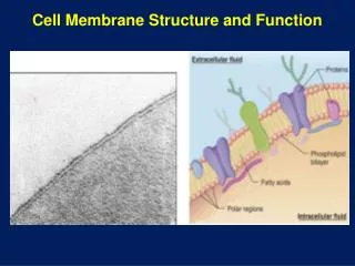
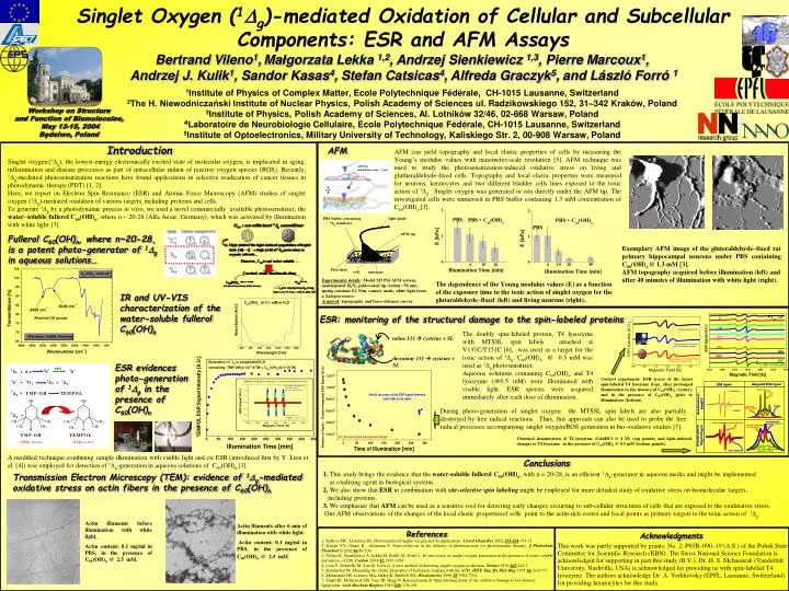
Singlet Oxygen (1Dg)-mediated Oxidation of Cellular and Subcellular Components: ESR and AFM Assays Bertrand Vileno1,Małgorzata Lekka 1,2, Andrzej Sienkiewicz 1,3, Pierre Marcoux1, Andrzej J. Kulik1, Sandor Kasas4, Stefan Catsicas4, Alfreda Graczyk5, and László Forró 1 1Institute of Physics of Complex Matter, Ecole Polytechnique Fédérale, CH-1015 Lausanne, Switzerland 2The H. Niewodniczański Institute of Nuclear Physics, Polish Academy of Sciences ul. Radzikowskiego 152, 31–342 Kraków, Poland 3Institute of Physics, Polish Academy of Sciences, Al. Lotników 32/46, 02-668 Warsaw, Poland 4Laboratoire de Neurobiologie Cellulaire, Ecole Polytechnique Fédérale, CH-1015 Lausanne, Switzerland 5Institute of Optoelectronics, Military University of Technology, Kaliskiego Str. 2, 00-908 Warsaw, Poland Workshop on Structure and Function of Biomolecules, May 13-15, 2004 Będelwo, Poland Introduction AFM AFM can yield topography and local elastic properties of cells by measuring the Young’s modulus values with nanometer-scale resolution [5]. AFM technique was used to study the photosensitization-induced oxidative stress on living and glutheraldehyde-fixed cells. Topography and local elastic properties were measured for neurons, keratocytes and two different bladder cells lines exposed to the toxic action of 1Dg. Singlet oxygen was generated in situ directly under the AFM tip. The investigated cells were immersed in PBS buffer containing 1.3 mM concentration of C60(OH)n [3]. Singlet oxygen(1Dg), the lowest-energy electronically excited state of molecular oxygen, is implicated in aging, inflammation and disease processes as part of intracellular milieu of reactive oxygen species (ROS). Recently, 1Dg-mediated photosensitization reactions have found applications in selective eradication of cancer tissues in photodynamic therapy (PDT) [1, 2]. Here, we report on Electron Spin Resonance (ESR) and Atomic Force Microscopy (AFM) studies of singlet oxygen (1Dg)-mediated oxidation of various targets, including proteins and cells. To generate 1Dg by a photodynamic process in vitro, we used a novel commercially available photosensitizer, the water–soluble fullerol C60(OH)n, where n ~ 20-28 (Alfa Aesar, Germany), which was activated by illumination with white light [3]. Fullerol C60(OH)n, where n~20-28, is a potent photo-generator of 1g in aqueous solutions… Exemplary AFM image of the gluteraldehyde–fixed rat primary hippocampalneurons under PBS containing C60(OH)n @ 1.3 mM [3]. AFM topography acquired before illumination (left) and after 40 minutes of illumination with white light (right). Experimental details: Model M5 PSI AFM system, unsharpened Si3N4 gold-coated tip (radius ~50 nm), spring constant 0.1 N/m, contact mode, white light from a halogen source. Acquired: topography and force-distance curves. The dependence of the Young modulus values (E) as a function of the exposure time to the toxic action of singlet oxygen for the glutaraldehyde–fixed (left) and living neurons (right). IR and UV-VIS characterization of the water-soluble fullerol C60(OH)n ESR: monitoring of the structural damage to the spin-labeled proteins The doubly spin-labeled protein, T4 lysozyme with MTSSL spin labels attached at V131C/T151C [6], was used as a target for the toxic action of 1Dg. C60(OH)n @ 0.5 mM was used as 1Dg photosensitizer. Aqueous solutions containing C60(OH)n and T4 lysozyme (@0.5 mM) were illuminated with visible light. ESR spectra were acquired immediately after each dose of illumination. valine 131 cysteine + SL threonine 151 cysteine + SL ESR evidences photo-generation of 1g in the presence of C60(OH)n Control experiment: ESR traces of the intact spin-labeled T4 lysozyme (top), after prolonged illumination in the absence of C60(OH)n (center), and in the presence of C60(OH)n prior to illumination (bottom). During photo-generation of singlet oxygen the MTSSL spin labels are also partially destroyed by free radical reactions. Thus, this approach can also be used to probe the free radical processes accompanying singlet oxygen/ROS generation in bio-oxidative studies [7]. Chemical denaturation of T4 lysozyme (GdnHCl @ 4 M) (top panels) and light-induced changes to T4 lysozyme in the presence of C60(OH)n@ 0.5 mM (bottom panels). A modified technique combining sample illumination with visible light and cw ESR (introduced first by Y. Lion et al. [4]) was employed for detection of 1g-generation in aqueous solutions of C60(OH)n [3]. Conclusions 1. This study brings the evidence that the water-soluble fullerol C60(OH)n, with n = 20-28, is an efficient 1g-generator in aqueous media and might be implemented as oxidizing agent in biological systems. 2. We also show that ESR in combination with site-selective spin labeling might be employed for more detailed study of oxidative stress on biomolecular targets, including proteins. 3. We emphasize that AFM can be used as a sensitive tool for detecting early changes occurring to sub-cellular structures of cells that are exposed to the oxidatative stress. Our AFM observations of the changes of the local elastic propertiesof cells point to the actin-rich cortex and focal points as primary targets to the toxic action of 1g. Transmission Electron Microscopy (TEM): evidence of 1g-mediated oxidative stress on actin fibers in the presence of C60(OH)n Actin filaments before illumination with white light. Actin content: 0.1 mg/ml in PBS, in the presence of C60(OH)n @ 2.5 mM. Actin filaments after 6 min of illumination with white light. Actin content: 0.1 mg/ml in PBS, in the presence of C60(OH)n @ 2.5 mM. References Acknowledgments