Download
1 / 54
550 likes | 582 Vues
Urinary System. The Urinary System. Function. Remove nitrogenous wastes Maintain electrolyte, acid-base, and fluid balance of blood Homeostatic organ Acts as blood filter Release hormones: calcitriol & erythropoietin. Kidneys as Filters. Diuretic- loose water; coffee, alcohol

E N D
Function • Remove nitrogenous wastes • Maintain electrolyte, acid-base, and fluid balance of blood • Homeostatic organ • Acts as blood filter • Release hormones: calcitriol & erythropoietin
Kidneys as Filters • Diuretic- loose water; coffee, alcohol • Antidiuretic- retain water; ADH • Aldosterone- sodium & water reabsorption, and K+ excretion • GFR= 180 liters (50 gal) of blood/day • 178-179 liters are reabsorbed back into blood • Excrete a protein free filtrate
The Urinary System Maintaining Chemical Homeostasis
blood filtration tubular reabsorption and secretion General Functioning of the Kidney “refreshed” blood urine
Nitrogenous Wastes urea uric acid ammonia
kidneys ureters urinary bladder urethra Organs of the Urinary System
renal pyramids renal pelvis renal cortex renal capsule ureter renal medulla Kidney Anatomy
nephron renal artery renal vein Kidney Anatomy
blood filtration tubular reabsorption and secretion Nephron Functioning “refreshed” blood urine
glomerulus efferent arteriole Bowman’s capsule afferent arteriole proximal convoluted tubule artery distal convoluted tubule peritubular capillaries vein collecting duct loop of Henle
Each kidney contains over 1 million nephrons and thousands of collecting ducts renal cortex renal medulla DCT Glomerulus PCT Collecting duct Loop of Henle
efferent arteriole afferent arteriole Bowman’s capsule glomerulus Glomerular Filtration Filters blood; proteins can’t pass through
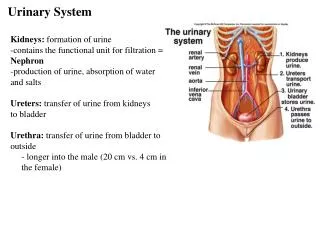
Radiographic examinations of the urinary system are among the most common contrast media procedures performed in radiology departments. The urinary system consists of two kidneys, two ureters, one urinary bladder, and one urethra
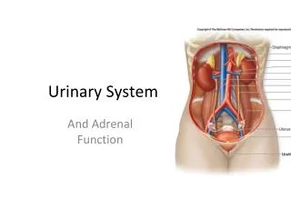
The two kidneys and the ureters are organs that lie in the retroperitoneal space. These two bean-shaped organs lie on either side of the vertebral column in the most posterior part of the abdominal cavity. The right kidney generally is slightly lower or more inferior than the left because of the presence of the liver. Near the upper medial part of each kidney is a suprarenal (adrenal) gland. These important glands of the endocrine system are located in the fatty capsule that surrounds each kidney. Each kidney is connected to the single urinary bladder by its own ureter. Waste material, in the form of urine, travels from the kidneys to the bladder via these two narrow tubes, termed ureters. The saclike urinary bladder serves as a reservoir that stores urine until it can be eliminated from the body via the urethra. The Latin designation for kidney is ren, and renal is an adjective that is commonly used to refer to the kidney.
Anterior view Posterior view
Kidneys the posteriorly placed kidneys lie on either side of the vertebral column in the upper posterior abdomen. They lie posterior to the lower portion of the liver on the right and posterior to the lower spleen on the left . The lower ribcage thus forms a protective enclosure for the kidneys. Ureters most of each ureter lies anterior to its respective kidney. The ureters follow the natural curve of the vertebral column. Each ureter initially curves forward, following the lumbar lordotic curvature, and then curves backward on entering the pelvis. After passing into the pelvis, each ureter follows the sacrococcygeal curve before entering the posterolateral aspect of the bladder The urethra connects the bladder to the exterior. The urethra exits from the body inferior to the symphysis pubis. The entire urinary system is either posterior to or below the peritoneum. The kidneys and ureters are retroperitoneal structures, whereas the bladder and urethra are infraperitoneal structures.
The usual orientation of the kidneys in the supine individual is shown below. The large muscles on either side of the vertebral column cause the longitudinal plane of the kidneys to form a vertical angle of about 20° with the midsagittal plane. These large muscles include the two psoasmajor muscles. These muscle masses grow larger as they progress inferiorly from the upper lumbar vertebrae. This gradual enlargement produces the 20° angle, wherein the upper pole of each kidney is closer to the midline than its lower pole
These large posterior abdominal muscles also cause the kidneys to rotate backward within the retroperitoneal space. As a result, the medial border of each kidney is more anterior than the lateral border. Transverse cross-sectional views through the level of L2 illustrate the usual amount of backward rotation of the kidneys.The normal kidney rotation of about 30° is due to the midline location of the vertebral column and the large psoas major muscles on either side. The quadratus lumborum muscles also are shown on each side just posterior to the kidneys. The deep muscles of the back include the group of erector spinae muscles on each side of the spine.
When posterior oblique projections are used during radiographic studies of the urinary system, each kidney in turn is placed parallel to the plane of the image receptor. The body is rotated about 30° in each direction to place one kidney, and then the other, parallel to the IR plane. A 30° LPO positions the right kidney parallel to the IR, and a 30° RPO positions the left kidney parallel.
Most abdominal radiographs are performed on expiration with the patient supine. The combined effect of expiration and a supine position allows the kidneys to lie fairly high in the abdominal cavity. Under these conditions, the kidneys normally lie about halfway between the xiphoid process and the iliac crest. The left kidney normally lies about 1 centimeter more superior than does the right one. The top of the left kidney is usually at the level of the T11-T12 interspace. The bottom of the right kidney most often is level with the upper part of L3
Because the kidneys are only loosely attached within their fatty capsule, they tend to move up and down with movements of the diaphragm and position changes. When one inhales deeply, the kidneys normally drop about 1 inch (2.5 cm) or one lumbar vertebra. When one stands upright, the kidneys normally drop about one lumbar vertebrae, or 5 centimeters (2 inches). If the kidneys drop farther than this, a condition termed nephroptosis is said to exist. With some very thin and older patients in particular, the kidneys may drop dramatically and end up within the pelvis, which may create problems caused by “kinking” or twisting of the ureters.
The primary function of the urinary system is the production of urine and its elimination from the body. During urine production, the kidneys perform the following functions: 1.Remove nitrogenous wastes 2.Regulate water levels in the body 3.Regulate acid-base balance and electrolyte levels of the blood Nitrogenous waste products such as urea and creatinine are formed during the normal metabolism of proteins. Buildup of these nitrogenous wastes in the blood results in the clinical condition termed uremia and may indicate renal dysfunction.
The macroscopic internal structure of the kidney.The outer covering of the kidney is termed the renal (fibrous) capsule. Directly under the renal capsule surrounding each kidney is the cortex, which forms the peripheral, or outer, portion of the kidney. Under the cortex is the internal structure termed the medulla, which is composed of from 8 to 18 conical masses termed renal pyramids. The cortex periodically dips between the pyramids to form the renal columns, which extend to the renal sinus.
The renal pyramids are primarily a collection of tubules that converge at an opening at the renal papilla (apex) and drain into the minor calyx. Calyces appear as hollowed, flattened tubes. From 4 to 13 minor calyces unite to form two to three major calyces. The major calyces unite to form the renal pelvis, which appears in the shape of a larger flattened funnel. Each expanded renal pelvis narrows to continue as the ureter. Thus urine formed in the microscopic or nephron portion of the kidney finally reaches the ureter by passing through the various collecting tubules, to a minor calyx, to a major calyx, and then to the renal pelvis. The general term renal parenchyma is used to describe the total functional portions of the kidneys, such as those visualized during an early phase of an intravenous urogram procedure. The structural and functional unit of the kidney is the microscopic nephron. Approximately one million nephrons exist within each kidney.. Small arteries in the kidney cortex form tiny capillary tufts, termed glomeruli(glo-mer′-u-li). Blood initially is filtered through the many glomeruli.
The ureters transport urine from the kidneys to the urinary bladder. Slow peristaltic waves and gravity force urine down the ureters into the bladder. This is an image taken 10 minutes after injection of contrast media into the bloodstream performed as part of an intravenous urogram procedure. The renal pelvis leaves each kidney at the hilum to become the ureter. The ureters vary in length from 28 to 34 centimeters, with the right one being slightly shorter than the left. As the ureters pass inferiorly, they lie on the anterior surface of each psoas major muscle. Continuing to follow the curvature of the vertebral column, the ureters eventually enter the posterolateral portion of each side of the urinary bladder.
The ureters vary in diameter from 1 millimeter to almost 1 centimeter. Normally, three constricted points exist along the course of each ureter. If a kidney stone attempts to pass from kidney to bladder, it may have trouble passing through these three regions. The first point is the ureteropelvic(u-re′-ter-o-pel′-vic)(UP) junction, where the renal pelvis funnels down into the small ureter. This section is best seen on the radiograph in. The second is near the brim of the pelvis, where the iliac blood vessels cross over the ureters. The third is where the ureter joins the bladder, termed the ureterovesicaljunction, or UV junction. Most kidney stones that pass down the ureter tend to hang up at the third site, the UV junction, and once the stone passes this point and moves into the bladder, it generally has little trouble passing from the bladder and through the urethra to the exterior.
The urinary bladder is a musculomembranous sac that serves as a reservoir for urine. The empty bladder is somewhat flattened and assumes the more oval shape only when partially or fully distended. The triangular portion of the bladder along the inner, posterior surface is termed the trigone . The trigone is the muscular area formed by the entrance of the two ureters from behind and the exit site of the urethra. The trigone is firmly attached to the floor of the pelvis. The mucosa of the trigone is smooth, whereas the remaining aspect of the inner mucosa of the bladder has numerous folds termed rugae. As the bladder fills, the top of the bladder expands upward and forward toward the abdominal cavity. The bladder functions as a reservoir for urine and, aided by the urethra, expels urine from the body. Normally, some urine is in the bladder at all times, but as the amount reaches 250 ml, the desire to void arises. The act of voiding (urination) is normally under voluntary control, and the desire to void may pass if the bladder cannot be emptied right away. The total capacity of the bladder varies from 350 to 500 ml. As the bladder becomes more and more full, the desire to void becomes more and more urgent. If the internal bladder pressure rises too high, involuntary urination occurs.
Venipuncture is defined as the percutaneous puncture of a vein for withdrawal of blood or injection of a solution such as contrast media for urographic procedures. In the past, venipuncture for urography was performed by physicians and laboratory or nursing personnel. However, in recent years, venipuncture has become part of the scope of practice for the diagnostic imaging professional. Before contrast media is withdrawn from any vial or bottle, confirmation of the correct contents of the container, route of administration, amount to be administered, and expiration date is imperative. Water-soluble, iodinated contrast media is used for radiographic examinations of the urinary system. This type of contrast medium can be administered by either bolus injection or drip infusion. A bolus injection is one in which the entire dose of contrast media is injected into the venous system at one time. This method of administration is used typically for maximum contrast enhancement.
Ch 26: Urinary System Objectives Identify and describe the components of the urinary system and their function Describe the (histological) organization of the nephron Identify the blood vessels that supply blood to the nephrons Describe the blood flow through and around the nephron
Functions of Urinary System (Kidneys): • Regulate fluid balance (fluid volume) of the body • Excrete organic waste products and conserve nutrients, etc • Stabilize pH • Regulate ion concentrations in the blood
Kidney Location Lateral to vertebral column high on body wall, under floating ribs in retro-peritoneal position
Surface Anatomy Size of bar of soap Bean shaped Hilus – indentation Three layers • Renal fascia – fibrous tunic • Adipose capsule – protects kidney • Renal capsule – anchors kidney to body wall, continuous with peritoneum
Sectional Anatomy Cortex:outer layer, light reddish brow, granular appearance (due to many capillaries) Medulla:darker striped appearance (due to tubules) Subdivided into distinct renal pyramids, terminating with a papilla. Separated by renal columns from the cortex. Fig 26-3
Urine collection: Ducts within each renal papilla release urine into minor calyx major calyx renal pelvis ureter
Segmental arteries Interlobar arteries Arcuate arteries Interlobular arteries Afferent arterioles Glomerulus Efferent arterioles Peritubular capillaries Segmental veins Interlobar veins Arcuate veins Interlobular veins Venules Renal Circulation Renal Artery Renal Vein Fig 26-4
Functional unit: Nephron (>mio/kidney) • Renal corpuscle: • Glomerulus • Bowman’s capsule • Tubular passageways with associated blood vessels: • PCT • LOH • DCT • CD Fig 26-8 Fig 26-6
Filtration: Passage across Three Barriers Fig 26-8 Capillary endothelium Fenestrated What gets through? Basement membrane Glomerular epithelium (= visceral layer of Bowman’s capsule) slit pores between pedicels of podocyte
Two Types of Nephrons • Cortical nephrons (85%) shorter, mostly in cortex of kidney, produce "standard" urine • Juxtamedullary nephrons (15%), "juxta-next-to" the medulla - responsive to ADH, can concentrate urine
Juxtaglomerular Apparatus Macula densa + Juxtaglomerular cells (smooth muscle fibers from afferent arteriole) = Juxtaglomerular Apparatus = Endocrine system structure (renin and EPO)
Urine Transport, Storage, and Elimination • Trace drop of urine from kidneys to outside world • Lining of these parts? • Nephroptosis (= floating kidneys) • Nephrolithiasis
Nephrolithiasis Occurs when urine becomes too concentrated and substances crystalize. Symptoms arise when stones begin to move down ureter causing intense pain. Kidney stones may form in the pelvis or calyces of the kidney or in the ureter.