Download
1 / 30
300 likes | 310 Vues
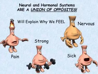
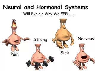
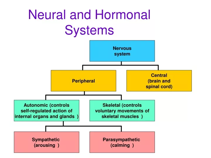
Nervous system. Peripheral. Central (brain and spinal cord). Autonomic (controls self-regulated action of internal organs and glands). Skeletal (controls voluntary movements of skeletal muscles). Sympathetic (arousing). Parasympathetic (calming). Neural and Hormonal Systems.

E N D
Nervous system Peripheral Central (brain and spinal cord) Autonomic (controls self-regulated action of internal organs and glands) Skeletal (controls voluntary movements of skeletal muscles) Sympathetic (arousing) Parasympathetic (calming) Neural and Hormonal Systems
Neurons and Synapses Types of Neurons Sensory/Afferent Motor/Efferent Interneurons
Sensory Neurons • INPUT Fromsensory organs to the brain and spinal cord. Brain Drawing shows a somatosensory neuron Vision, hearing, taste and smell nerves are cranial, not spinal Sensory Neuron Spinal Cord
Brain Sensory Neuron Spinal Cord Motor Neuron Motor Neurons • OUTPUTFrom the brain and spinal cord To the muscles and glands.
Brain Sensory Neuron Spinal Cord Motor Neuron Interneurons • Interneurons carry information between other neurons only found in the brain and spinal cord.
The cell body • Round, centrally located structure • Contains DNA • Controls protein manufacturing • Directs metabolism • No role in neural signaling • Contains the cell’s Nucleus
Dendrites • Information collectors • Receive inputs from neighboring neurons • Inputs may number in thousands • If enough inputs the cell’s AXON may generate an output
Dendritic Growth • Mature neurons generally can’t divide • But new dendrites can grow • Provides room for more connections to other neurons • New connections are basis for learning
Axon • The cell’s output structure • One axon per cell, 2 distinct parts • tubelike structure branches at end that connect to dendrites of other cells
Myelin Sheath Myelin sheath • White fatty casing on axon • Acts as an electrical insulator • Not present on all cells • When present increases the speed of neural signals down the axon.
How neurons communicate • Neurons communicate by means of an electrical signal called the Action Potential • Action Potentials are based on movements of ions between the outside and inside of the cell • When an Action Potential occurs a molecular message is sent to neighboring neurons
Outside of Cell K+ Na+ Cl- Cell Membrane in resting state K+ Na+ Cl- A- Inside of Cell Ion concentrations
K+ Na+ Cl- Outside of Cell Cell Membrane at rest Na+ - 70 mv A- K+ Cl- Inside of Cell Potassium (K+) can pass through to equalize its concentration Sodium and Chlorine cannot pass through Result - inside is negative relative to outside The Cell Membrane is Semi-Permeable In a resting state – there is more negatively charged molecules on the inside than outside & more positively charged ions on the outside than inside.
Resting Potential • At rest the inside of the cell is at -70 microvolts • With inputs to dendrites the inside becomes more positive • if resting potential rises above threshold an action potential starts to travel from cell body down the axon • Figure shows resting axon being approached by an AP
Depolarization ahead of AP • AP opens cell membrane to allow sodium (NA+) in • inside of cell rapidly becomes more positive than outside • this depolarization travels down the axon as leading edge of the AP
Repolarization follows • After depolarization potassium (K+) moves out restoring the inside to a negative voltage • This is called repolarization • The rapid depolarization and repolarization produce a pattern called a spike discharge
Finally, Hyperpolarization • Repolarization leads to a voltage below the resting potential, called hyperpolarization • Now neuron cannot produce a new action potential • This is the refractory period
Dendrite Axon Cell Body Neuron to Neuron • Axons branch out and end near dendrites of neighboring cells • Axon terminals are the tips of the axon’s branches • A gap separates the axon terminals from dendrites • Gap is the Synapse
Sending Neuron Axon Synapse Terminal Synapse • axon terminals contain small storage sacs called synaptic vesicles • vesicles contain neurotransmitter molecules
Neurotransmitter Release • Action Potential causes vesicle to open • Neurotransmitter released into synapse • Locks onto receptor molecule in postsynaptic membrane
Locks and Keys • Neurotransmitter molecules have specific shapes • Receptor molecules have binding sites • When NT binds to receptor, ions enter positive ions (NA+ ) depolarize the neuron negative ions (CL-) hyperpolarize
Some Drugs work on receptors • Some drugs are shaped like neurotransmitters • Antagonists : fit the receptor but poorly and block the NT • e.g. beta blockers • Agonists : fit receptor well and act like the NT • e.g. nicotine. Website for neural simulations
Neurotransmitter molecule Receiving cell membrane Agonist mimics or enhances Neurotransmitter Receptor site on receiving neuron Antagonist blocks neurotransmitter Neural Communication Amphetamines & cocaine are agonists increasing dopamine in the brain Caffeine increases the availability of glutamate (keeps NS aroused) Alcohol is an agonist by increasing sensitivity of receptor site to the inhibitory GABA anti-depressants are agonists that increase the levels of norepinephrine and serotonin by blocking reuptake Curare is a antagonist for Ach Anti-psychotic drugs block dopamine in schizophrenics
The Endocrine System The endocrine system refers to a set of glands that produce chemical messengers called hormones.
The Body’s “Slow but Sure” Endocrine Message System • The endocrine system sends molecules as messages, just like the nervous system, but it sends them through the bloodstream instead of across synapses. • These molecules, called hormones, are produced in various glands around the body. • The messages go to the brain and other tissues.
produce hormones such as adrenaline/epinephrine, noradrenaline/norepinephrine, and cortisol. Adrenal Glands Adrenal Glands • The sympathetic “fight or flight” nervous system responds to stress by sending a message to adrenal glands to release the hormones listed above. • Effect: increased heart rate, blood pressure, and blood sugar. These provide ENERGY for the fight or flight! Pancreas
The Pituitary Gland • The pituitary gland is the “master gland” of the endocrine system. • It is controlled through the nervous system by the nearby brain area--the hypothalamus. • The pituitary gland produces hormones that regulate other glands such as the thyroid. • It also produces growth hormone (especially during sleep) and oxytocin, the “bonding” hormone. Pituitary gland