Download

1 / 45
450 likes | 1.28k Vues
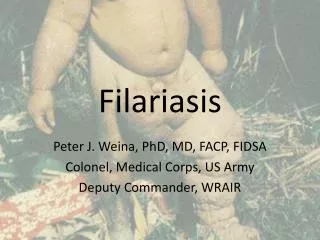
FILARIASIS. DEFINISI FILARIASIS. Filariasis ( philariasis ) is a parasitic disease caused by thread-like nematodes (roundworms) belonging to the superfamily Filarioidea , [1] also known as " filariae ". [2] These are transmitted from host to host by blood-feeding arthropods ,

E N D
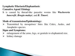
DEFINISI FILARIASIS Filariasis (philariasis) is a parasitic disease caused by thread-like nematodes (roundworms) belonging to the superfamily Filarioidea,[1] also known as "filariae".[2] These are transmitted from host to host by blood-feeding arthropods, mainly black flies and mosquitoes.
JENIS FILARIASIS (menurutlokasiinfeksi). 1.Lymphatic filariasis 2.Subcutaneous filariasis 3.Serous cavity filariasis
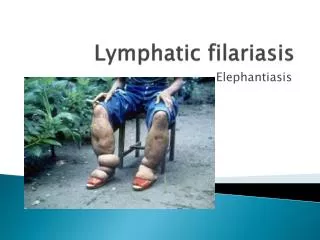
JENIS FILARIASIS (menurutlokasiinfeksi). 1.Lymphatic filariasis is caused Wuchereriabancrofti, Brugiamalayi, and Brugiatimori. In the lymphatic system, the lymph nodes, in chronic cases lead to elephantiasis. 2.Subcutaneous filariasis is caused by Loa loa (the eye worm), Mansonellastreptocerca, and Onchocerca volvulus. In the subcutaneous layer of the skin, in the fat layer. L. loa causes Loa loafilariasis O. volvulus causes river blindness. 3.Serous cavity filariasis is caused by Mansonellaperstans and Mansonellaozzardi, in the serous cavity of the abdomen.
VECTOR FILARIA W. bancroftiperkotaan culexquinquefasciatus W. bancroftipedesaan: anopheles, aedesdanarmigeres B. malayi : mansoniaspp, an.barbirostris. B. timori : an. barbirostris
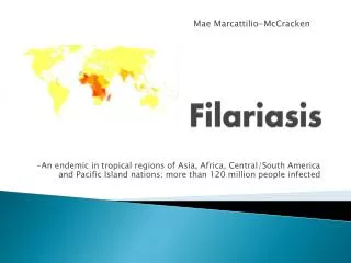
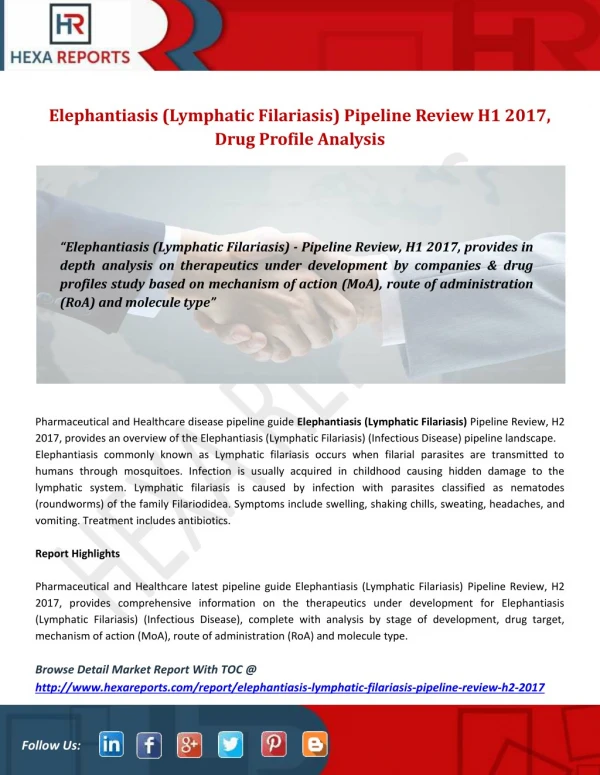
EPIDEMIOLOGY International 1.Lymphatic filariasis 90 million people and throughout the tropics and subtropics INFECTED. 2.O volvulus in equatorial Africa and foci in Central and South America. At least 21 million people INFECTED. 3. L loa Approximately 3 million people in Central Africa are infected . In 1997, the World Health Organization (WHO) initiated a program to globally eliminate lymphatic filariasis as a public health problem.
EPIDEMIOLOGY Mortality/Morbidity Filarial diseases are rarely fatal, infection can cause significant personal and socioeconomic hardship The WHO has identified lymphatic filariasis as the second leading cause of permanent and long-term disability in the world after leprosy. The morbidity of human filariasis mainly from the host reaction to microfilariae or developing adult worms in different areas of the body.
EPIDEMIOLOGY Race Filariasis has no known racial predilection. Sex Both sexes are equally susceptible to filariasis. Age All ages are susceptible and potentially microfilaremic. Microfilaremia rates increase with age through childhood and early adulthood, although clinical infection may not be apparent. The manifestation of acute and chronic filariasis usually occurs only after years of repeated and intense exposure to infected vectors in endemic areas.
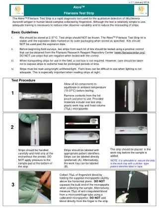
LYMPHATIC FILARIASIS Disease caused by nematode worms of the genera WucheriaandBrugia. Larval worms circulate in the bloodstream of infected persons, and adult worms live in the lymphatic vessels. Screening. Blood sample collected in the middle of the night with the time of peak microfilariae abundance. ELISA test for antigens of the parasite in blood samples collected any time of the day is now available, easier.
KRITERIA ENDEMIS /PENULARAN FILARIASIS Kriteria penularan penyakit mikrofilarial rate ≥ 1% pada sample darahdi sekitarkasus elephantiasis, atauadanya 2 ataulebihkasus elephantiasis di suatuwilayahpadajarakterbangnyamukyang mempunyairiwayatmenetapbersama/berdekatan padasuatuwilayahselamalebihdarisatutahun. Berdasarkan WHO, mikrofilarial rate ≥ 1%pada satuwilayahmakadaerahtersebutdinyatakan endemis ,harusdiberikanpengobatanmasal selama5 tahunberturut-turut.
GEJALA PENYAKIT FILARIASIS 1. gejala dan tanda klinis akut : Demamberulangulangselama 3-5 hari, demamdapathilangbilaistirahatdan timbullagisetelahbekerjaberat Pembengkakankelenjargetahbening (tanpaadaluka) di daerahlipatanpaha, ketiak (limfadenitis) yang tampakkemerahan, panasdansakit Radangsalurankelenjargetahbening yang terasapanasdansakit yang menjalar daripangkalkearahujung kaki ataulengan Absesfilaria terjadiakibatseringnyapembengkakankelenjargetahbening, dapatpecahdandapatmengeluarkandarahsertananah Pembesarantungkai, lengan, buah dada danalatkelaminperempuandanlaki-laki yang tampakkemerahandanterasapanas.
GEJALA PENYAKIT FILARIASIS 2. Gejala dan tanda klinis kronis : Limfedema : InfeksiWuchereriamengenai kaki danlengan, skrotum, penis, vulva vagina danpayudara, InfeksiBrugiadapatmengenai kaki danlengan dibawahlutut / siku lututdansikumasih normal Hidrokel: Pelebarankantungbuahzakar yang berisicairanlimfe, dapatsebagai indikatorendemisitasfilariasisbancrofti Kiluria :Kencingsepertisusu kebocoransellimfe di ginjal, j
DIAGNOSA FILARIASIS DIAGNOSIS FILARIASIS Klinis diagnosis klinisditegakkanbiladitemukangejaladantandaklinisakutataupunkronis 2. Laboratorium Dinyatakan sebagai penderita falariasis apabila dalam darahnyapositifditemukanmikrofilaria. Darah jari yang diambil pada malam hari (pukul 20.00 - 02.00). Test ELIZA tidak perlu malam hari
PENGOBATAN 1. PengobatanMasal di daerah endemis (mf rate > 1%) Diethyl CarbamazineCitrate (DEC) dikombilansikanAlbendazole sekalisetahunselama 5 tahunberturut-turut. Pengobatanmassalseluruh pendudukyang usia > 2 tahun Ditundausia≤ 2 tahun, wanitahamil, ibumenyusui
PENGOBATAN 2. PengobatanSelektif Dilakukan pada orang yang mengidap mikrofilaria anggota keluarga yang tinggal serumah danberdekatandenganpenderita (hasilsurvey mikrofilaria<1% (non endemis) 3. Pengobatan Individual (penderitakronis) Semua kasus klinis diberikan obat DEC 100 mg, 3x sehari selama 10 hari sebagai perawatanterhadaporgan yang bengkak
SYMPTOMATOLOGY 1.Asymptomatic : 70 % are asymptomatic. Symptoms usually do not manifest until adolescence or adulthood, when worm burden is usually the highest.
Lymphatic filariasis The symptoms of lymphatic filariasis predominantly result from the presence of adult worms residing in the lymphatics. The clinical course is broadly divided into 1.asymptomatic microfilaremia, 2.acute phases of adenolymphangitis (ADL), 3.chronic irreversible lymphedema.
Three acute syndromes in filariasis, as follows: 1.Acute ADL: This refers to the sudden onset of febrile painful lymphadenopathy. Pathologically, the lymph node is characterized by a retrograde lymphangitis, distinguishing it from bacterial lymphadenitis. Symptoms usually abate within one week, but recurrences are possible. 2.Filarial fever: characterized by fever without the associated adenitis. 3.Tropical pulmonary eosinophilia (TPE)
Tropical pulmonary eosinophilia TPE is a form of occult filariasis. Presenting symptoms include a paroxysmal dry cough, wheezing, dyspnea, anorexia, malaise, and weight loss. Symptoms of TPE are usually due to the inflammatory response to the infection. Characteristically, peripheral blood eosinophilia and abnormal findings on chest radiography are observed. TPE is usually related to W bancrofti or B malayi infection.
Onchocerciasis This also is known as hanging groins, leopard skin, river blindness, or sowda. Symptoms Microfilariae in the skin and include pruritus, subcutaneous lumps, lymphadenitis, and blindness. Patients with onchocerciasis may report impaired visual acuity due to corneal fibrosis.
Loiasis The symptoms Lloa infection are usually to subcutaneous swellings on the extremities, localized pain, pruritus, and urticaria. Microfilaremia tends to be asymptomatic. Occasionally, the worm is observed migrating through the subconjunctiva or other tissues.
M ozzardi, M perstans, and M streptocerca • Mansonella infections are usually asymptomatic. • If symptoms are present, • fever, pruritus, skin lumps, • lymphadenitis, and abdominal pain. MANSONIAMOSQUITO