Download
1 / 24
340 likes | 831 Vues
University of Hail Faculty of Sciences Department of Biology. Practical Biology Biol 101 Lab 4. Biological Macromolecules. All living organisms are composed of four classes of macromolecules: Carbohydrates Lipids Proteins Nucleic acid.

E N D
University of Hail Faculty of Sciences Department of Biology Practical Biology Biol 101Lab 4 Biological Macromolecules
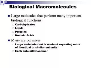
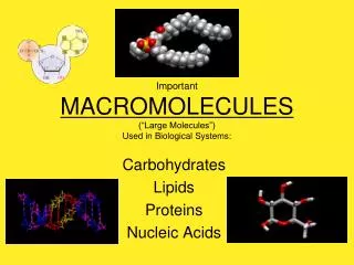
All living organisms are composed of four classes of macromolecules: • Carbohydrates • Lipids • Proteins • Nucleic acid
Carbon containing compounds are called organic compounds. • Carbon is the backbone of organic molecules, but its only one of several important bioelements. • These bioelements are hydrogen, nitrogen, oxygen, phosphorous and sulfur.
The chemical properties of the different classes depend on the presence of specific functional groups: • Aldehyde group • Ketone group • Carboxyl group • Amino group
Identification of Carbohydrates • Carbohydrates are the main energy storing molecules. • Monosaccharide : monomers (glucose, ribose, galactose). • Disaccharides: two monomers connected with glycosidic bond (sucrose, lactose). • Polysaccharides: polymers; more than three monomers (starch).
Identification of carbohydrates A. Benedict’s test for reducing sugars: • The test depends on the presence of free Aldehyde or ketone group. • Monosaccharides and some disaccharides have these groups free are called reducing sugars. • Free groups found in reducing sugars will react with Benedict’s reagent (CuSo4 which is blue in color with NaOH). • The test is both qualitative and quantitative test; • Small amount of reducing sugar green color. • Large amount of reducing sugar red- orange color.
Procedure: • Label 7 clean test tubes • (1-7) • Fill the test tubes with 1 ml of one of the following solutions. apple juice glucose sucrose D.W maltose starch milk
Fill each test tube with 3 ml of Benedict’s reagent • Put the test tubes in boiling water for 5 minutes • If color change to: • Yellow • Orange Positive test for reducing. • Red • Green • If color remains blue • (the color of Benedict’s reagent) Negative result. • Record your results in a proper table
apple juice glucose sucrose maltose D.W starch milk
B. Lugol’s iodine test for starch • Starch is a polysaccharide consisting of many glucose monomers linked together into long branching chains. • It is the primary storage carbohydrate in plants. • In the presence of iodine (I2-KI), a solution containing starch will turn blue-black in color.
Procedure: • Label 7 clean test tubes • (1-7) • Fill the test tubes with 2 ml of one of the following solutions. Potato juice apple juice glucose sucrose D.W starch milk 11
Add two drops of lugol’s iodine to each test tube. • If color change to: Positive test for starch • A blue-black • If color remains yellow - brown (the color of the iodine) Negative result. • Record the result in the report.
Potato juice apple juice glucose sucrose milk starch D.W 13
Identification of Proteins • Proteins are the key substances in the structural and physiological function of living things. • Proteins are polymers of amino acids in which the carboxyl group of one amino acid is linked with the amino group of the next amino acid in a covalent bond called the peptide bond. A. Ninhydrin test for amino acids • Ninhydrin reagent reacts with free amino groups i.e free amino acids to form a purple or violet colored substance.
Ninhydrin reagent can also be used to detect proteins, but they must be heated or digested to hydrolyze the protein into free amino acids. Procedure • label 4 clean test tubes (1-4) • Fill the test tubes with 2 ml of one of the following solutions: • Albumin solution. • Milk. • Amino acid solution (Lysine). • Distilled water.
To each of the test tubes, add 10 drops of ninhydrin reagent and heat the test tubes in boiling water for 5 minutes (avoid inhaling, poisonous fumes). • A purple color is the +ve result. • Record the color of the tubes content in the lab report.
B. Biruet test for polypeptides • The test reveals the presence of peptide bond i.e. proteins • Biuret reagent CuSo4 reacts with the peptide bonds between the amino acids changing in color from light blue to violet under alkaline conditions. • The intensity of the violet color is proportional to the protein concentration. • In the test, Cu+2must complex with at least four to six peptide bonds to produce a color. • Biuret reagent does not react with free amino acids. • Short peptides don’t react positively.
Procedure: • Label 5 clean test tubes • (1-5) • Fill the test tubes with 2 ml of one of the following solutions. D.W albumine Lysine starch milk 18
Fill each test tube with 2 ml of Biuret reagent • If color change to: • Violet Positive test for Biuret test • If color remains blue (the color of Biuret reagent). Negative result. • Record your results in a proper table. 19
albumine D.W starch Lysine milk
Identification of lipids • Lipids are a heterogeneous group of compounds that are insoluble in water, but are soluble in organic solvents such as ether and acetone. • There are three major classes of lipids: • Neutral fats (triglycerides). • Phospholipids • Steroids A. Sudan red test for fats • Sudan red is a lipid soluble dye, when added to a mixture of lipids and water, The dye will move into the lipid layer giving the lipid a red color.
Procedure • Fill the test tube with 2ml of Water . • Add 10 drops of oil and mix. • The two liquids do not mix after shaking the test tube, because the oil molecules are hydrophobic (insoluble in water) to produce an emulsion.
Add 10 drops of Sudan IV solution to the test tube and mix. • Two layers are formed, the upper one is the lipid stained with Sudan red IV • The water remains in the bottom layer • Record your observations.