Download
1 / 39
390 likes | 689 Vues
5 . X-ray spectroscopy. AIT. What are X-rays?. electromagnetic radiation very short wavelengths, meaning high frequencies and high energies wavelength range of the X‑ray region is 0.01 to 10 nm, compared to 400‑800 nm for the visible region up to 80000 x more energetic than visible radiation

E N D
What are X-rays? • electromagnetic radiation • very short wavelengths, meaning high frequencies and high energies • wavelength range of the X‑ray region is 0.01 to 10 nm, compared to 400‑800 nm for the visible region • up to 80000 x more energetic than visible radiation • an older unit ‑ the Angstrom ‑ is often used in measures of wavelength • 1Å is equal to 0.1 nm • X-ray range is 0.1 to 100 Å • keV also used – measure of energy • 1 nm = 1.24 keV; range from 124 to 0.124 keV (inverse)
X‑rays are produced in three ways: • when high energy electrons collide with a surface • loss of energy produces a continuous band of X‑ray wavelengths (used in X-ray sources) • emissions from decaying radioactive nuclei • also used in X-ray sources • emission of X‑rays from matter which has been irradiated with a X‑ray beam • single wavelengths, and of different energies to the excitation beam (X-ray fluorescence)
Safety aspects • high energies of X‑rays mean that they are potentially harmful to living organisms • the energy will be absorbed by molecules in the organism • cause destruction or alteration to the molecules absorbing the energy • direct • the photon breaks bonds in an important molecule • indirect • the photon splits water, forming the very reactive OH radical • this reacts with something important
Safety aspects • if the species affected is genetic material (DNA) • chance exists for mutated cells to be produced • leading to the formation of cancers • effect is cumulative • a limited number of X‑rays that a person can have with in a set period of time • too many increases the number of damaged molecules • the chance of serious health consequences
Safety aspects • instruments using them are thoroughly sealed so that no radiation should escape • seals are checked on a regular basis • removable/opening panels have micro-switches • trip out the X‑ray source within milliseconds • operators of X‑ray instruments wear radiation badges • developed each month to check the levels of exposure • still appear to be film-based Why won’t we ask you to wear one? • one or two pracs are not enough potential exposure
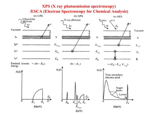
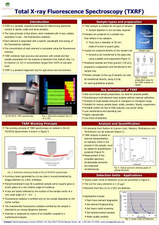
X‑ray spectroscopic techniques X‑ray diffraction (XRD) • where X‑rays are diffracted through characteristic angles by the crystal structure of the matter • allows distinction between different forms of the same element, e.g. between the various crystal structures that occur in steel, X‑ray fluorescence (XRF) • emission of characteristic wavelength X‑rays from matter irradiated by an X‑ray beam is used to identify and quantify particular elements X‑ray absorption (XRA) • the familiar use of X‑rays in medicine and materials testing • the varying density of matter causes variation in the absorption of the X-ray beam • only XRF part of this course
A bit of XRF history • 1895 - the discovery of X-rays is also the (not realised) discovery of XRF • 1913 – emitted X-rays are shown to be characteristic of different elements and related to atomic number • helps fix some errors in the periodic table • 1928 – first use of X-rays to generate XRF for analysis of real samples; limited by other instrumental problems • 1948 – first true working XRF spectrometer • 1950s – first commercial instruments • 1970 – use of semiconductor detection
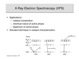
X-ray fluorescence • emission technique • released X-rays of lower energy than those absorbed • X-rays are energetic enough to penetrate to the inner electron orbitals and eject an electron completely from the atom • this creates a “hole” which the atom will fill by electrons from higher orbitals dropping down • in doing so they release energy • some of this energy is in the form of X-rays
M shell M & L electrons drop down L shell X-rays emitted X-ray K electron ejected K shell X-ray fluorescent transitions
X-ray transitions: • L K • MK • ML • NL • K, L, M and N shells are the old name for the 1st, 2nd, 3rd and 4th orbitals
Transition labels • transitions in XRF are given labels which identify them when a spectrum is recorded • labels indicate: • the shell from which the electron was ejected • K, L (change to notes: M rare, N no) • the shell from which the filling electron came • Greek letters , , and • the filling electron came from 1, 2 or 3 shells respectively, from the hole
M shell M & L electrons drop down L shell X-rays emitted X-ray K electron ejected K shell Exercise 5.2 (a) What are the two transitions shown in Figure 5.1? L K
N M L K Exercise 5.2 (b) Draw transitions for K and L. L K
Why are these labels important? • important trends in wavelength and intensity that can be followed for these transitions • help identify the presence of species in a sample • shorthand way of referring to certain characteristic emissions
N M L K Exercise 5.3 • Explain the order of energies for the four transitions • the bigger the jump, the greater the energy L La Ka K
Figure 5.2 Why use anything other than K? Not all X-rays can be excited efficiently or detected
Instrumentation • two basic designs of XRF instruments: • wavelength‑dispersive • the older type • uses a conventional monochromator-based design • energy‑dispersive • uses a special detector which doesn't require a monochromator
sample holder emitted radiation single detector path many s excitation X-rays detector collimator dispersing crystal X-ray source Wavelength dispersive some have moving crystal & fixed detector
Wavelength dispersive • in high resolution WD-XRFs both the detector & the diffracting crystal move around a circular path • this is known as the goniometer • this is the same as for the ICP • a large radius circular path gives better resolution than a rotating crystal through an exit slit
Energy dispersive X-ray source no monochromator! excitation X-rays detector emitted radiation sample collimator
Energy dispersive • no moving parts => able to be made portable • no scanning => instant spectrum • lower resolution => lesser ability to distinguish close wavelengths • lower sensitivity
window X-rays target electrons cathode Instrumental components – radiation source • an X‑ray tube consists of a heated wire cathode, which emits electrons • accelerated towards to the anode ‑ a block of metal known as the target • collision releases energy in the form of a continuous spectrum of X‑rays
Output variables for X-ray tube • applied voltage between the electrodes • current • the material used on the surface of the target
Other aspects • targets are water-cooled copper block with a surface coating of the target element • ours has a rhodium (Rh) target • the target element adds a number of X-ray lines to the continuous spectrum • alternative sources to the high current (30-50 kV) standard are: • radioactive isotopes (no power required) • low current X-ray tubes (portable or small desktop) Exercise 5.5 What would be the main disadvantage of these low current X-tubes? • lower current => lower intensity => lower fluorescence
The importance of tube voltage • voltage determines the energy of the exciting photons • => which elements will be excited. • the keV energy unit for X-rays is related to the voltage needed to excite a particular element line • to excite the fluorescence to generate that X-ray typically requires a tube voltage twice the keV value Example 5.1 The K line of calcium has an energy of 3.69 keV. What tube voltage needed to excite this line? • 2 x 3.69 = 7.38 kV
Exercise 5.6 The X-ray tube in our instrument has a maximum voltage of 30kV. What is the maximum atomic number for an element that can have its K line excited? • 30 ÷ 2 = 15 keV
Detectors • three main types: • semiconductor • gas-filled/flow/proportional • scintillation • a is used in energy-dispersive • b & c are used together in wavelength-dispersive • neither covers the entire range
Detectors Semiconductors • produce an output (a current pulse) that is proportional to the energy of the incoming photon • can do this for polychromatic radiation equally well • the number of pulses of each amount of current (e.g. 1, 1.1, 1.2 uA) gives the spectrum • correlation between energy and current must be made by some internal setting in the detector • eg 1.2 uA = 2.5 keV
Detectors - semiconductors • silicon-drift • do not need a monochromator • all the multi-channel-type advantages • limitations • resolution • sensitivity
Detectors Proportional/flow counters • X-rays ionise argon atoms • electrons generated can ionise other atoms, producing a chain reaction • this generates a current related to the number of photons • proportional relates to region where applied voltage is proportional to intensity • flow because of the throughput of gas • only measures lighter elements (higher wavelengths)
Detectors Scintillation counters • the generation of multiple photons of visible light from matter, which has absorbed a higher energy photon, e.g. an X‑ray • some crystalline materials do this • one X-ray can generate thousands of visible photons • detected by a photomultiplier tube • best for heavier elements (shorter wavelengths) • works in conjunction with flow counter to give full spectrum
Qualitative analysis XRF analysis: • is non-destructive • works better with solids • is of medium sensitivity (10 mg/kg upwards) • a surface technique • does not distinguish between different forms of an element • case studies • Victoria crosses • archaeological artefacts • medieval painting
Quantitative analysis • can quantitatively analyse solids • substantial matrix interferences • peak position is the same, but intensity varies greatly • sources of interference include: • preferential absorption of the excitation radiation • absorption of the analyte fluorescence • emission by matrix components • particle size and homogeneity variations
Means of countering matrix errors • matrix‑matched standards • standard addition • internal standard • standardless
Sample prepration • physical state affects intensity • equivalent treatment for standards and samples • solids need cleaning • powders need grinding & pressing or borax fusion
Other matters Use of helium • used for elements lighter than calcium for best sensitivity • low energy photons generated by these elements absorbed by air Filters • placed between tube and sample • absorb a particular range of excitation X-rays • tube lines can be removed by this method • loss of sensitivity Current control • a current that is too high will generate a large broad background “bump” in the spectrum • particularly if the sample has high concentrations of light elements