Download
1 / 34
340 likes | 527 Vues
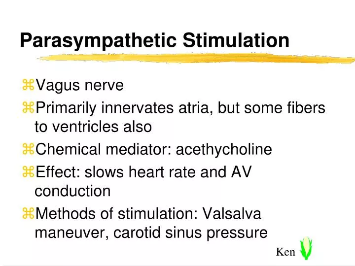
Parasympathetic Stimulation. Vagus nerve Primarily innervates atria, but some fibers to ventricles also Chemical mediator: acethycholine Effect: slows heart rate and AV conduction Methods of stimulation: Valsalva maneuver, carotid sinus pressure. Sympathetic Stimulation.

E N D
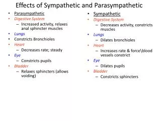
Parasympathetic Stimulation • Vagus nerve • Primarily innervates atria, but some fibers to ventricles also • Chemical mediator: acethycholine • Effect: slows heart rate and AV conduction • Methods of stimulation: Valsalva maneuver, carotid sinus pressure
Sympathetic Stimulation • Nerves arising in thoracic and lumbar ganglia • Innervate both atria and ventricles • Chemical mediator: norepinephrine • Receptor sites: alpha, beta
Effect of alpha Stimulation: • No effect on heart • Peripheral vasoconstriction
Effect of beta Stimulation: • Increased rate and conduction • Increased contractility • Bronchodilation • Peripheral vasodilation
Role of Electrolytes • Cardiac function, electrical and mechanical, influenced by electrolyte imbalances • Major electrolytes influencing cardiac function • Na+ Sodium • Ca++ Calcium • K+ Potassium
Role of Electrolytes • Sodium (Na +): major role in depolarization phase of myocardial cells • Calcium (Ca ++): major role in depolarization phase of myocardial pacemaker cells and in myocardial contractility • Hypercalcemia: increased myocardial contractility • Hypocalcemia: decreased myocardial contractility and increased electrical irritability
Role of Electrolytes • Potassium (K +): major role in repolarization phase • Hyperkalemia: decreased automaticity and conduction • Hypokalemia: increased irritability • Potassium levels are critical to life • Hyperkalemia = Tall peaked T waves
Electrophysiology • Electrical properties of the heart • Automaticity: ability to generate an electrical impulse without stimulation from another source ‑ property of pacemaker cells • Excitability: ability to respond to an electrical stimulus ‑property of all myocardial cells • Conductivity: ability to propagate an impulse from cell to cell
Allows electrical impulses to spread through the heart six times faster than through muscle alone Sequence of normal electrical conduction SA node Internodal and interatrial tracts AV node Bundle of His Bundle branches Purkinje fibers Electrical Conduction System
Function of electrical conduction structures • Sinoatrial (SA) node • Located in right atrium near entrance of superior vena cava • Usually heart's dominant pacemaker sa
Internodal and interatrial tracts • Pathways that carry impulse between SA node and AV node and spread it across atrial muscle • Impulse travel time: 0.08 seconds
Atrioventricular (AV) node: • Part of area called the "AV junctional tissue" along with some surrounding tissue and the non-branching portion of the Bundle of His • Responsible for creating slight delay in conduction before sending impulse to ventricles • Impulse travel time: 0.08‑0.16 seconds • No pacemaking properties in node itself
Bundle of His • Bundle of fibers coming off AV node, located at top of interventricular septum • Considered part of the AV junction • Makes electrical connection between atria and ventricles
Bundle branches • Created by bifurcation of Bundle of His into right and left branches • Carry electrical impulse at high velocity to interventricular septum and each ventricle simultaneously
Purkinje fibers • Terminal ends of bundle branches • Network of fibers helping to spread impulse throughout ventricular walls • Rapid impulse spread through ventricles: 0.08-0.09 seconds
Depolarization • Process by which muscle fibers are stimulated to contract by the alteration of electrical charge of the cell accomplished by changes in electrolyte concentrations across the cell membrane
Depolarization at The Cellular Level • Chemical pumps in cell wall maintain certain concentrations of electrolytes within and outside the cell • Resting (polarized) cell normally more electrically negative inside cell wall than outside ( -90 millivolts (mv) in working cells)
Depolarization at The Cellular Level • Electrical stimulation of cell wall changes its permeability to sodium (Na+) • Na+ rushes into cell, causing inside to become more positive • Slower influx of calcium (Ca++) also causes cell to become positive • Muscle contraction is response to depolarization • Depolarization wave is passed from cell to cell along the conduction pathway to reach the muscle cells
Spontaneous diastolic depolarization of pacemaker cells • Pacemaker cells capable of self-initiated depolarization (automaticity) • Found throughout conduction system except in AV node • During diastole, become less and less negative until a certain threshold reached, then rapidly and fully depolarize
Pacemaker Capabilities & Rates • SA node: 60-100/minute intrinsic rate • AV junctional tissue: 40-60/minute intrinsic rate • Ventricles (bundle branches and Purkinje fibers): 20-40/minute intrinsic rate • SA node usual pacemaker because it discharges the fastest; pacemaker cells below SA node normally suppressed by it
Repolarization • Process by which cells re-establish internal negativity and are readied for stimulation return to resting or polarized state • Caused by rapid escape of potassium (K+) from the cell • Proper distribution of electrolytes re-established by cell wall pumps (Na+ pumped out of cell, potassium pumped back into cell) • Cell returns to -90mv. internal charge- repolarized
Relationship of ECG to electrical activity • ECG is record of electrical activity of heart as sensed by electrodes on body surface • Gives information only about electrical activity tells us nothing about pump function • Isoelectric line: a flat line on the ECG indicating absence of net electrical activity
P wave • Rounded wave preceding QRS; usually upright (positive) in Lead II • Indicates depolarization of atrial muscle
QRS complex • Collective term for three deflections following the P wave
QRS complex • Wave-first negative deflection after P wave • R wave-first positive deflection after P wave • S wave-first negative deflection after R wave
QRS complex • All three waves not always present - QRS has many shapes • Indicates depolarization of the ventricular muscle
T wave • Rounded wave following QRS complex; usually in same direction as QRS • Indicates repolarization of ventricles • Atrial T wave (atrial repolarization) usually not visible buried within QRS complex
P-R interval • Distance between beginning of P wave and the beginning of QRS complex • Indicates length of time it takes depolarizatin wave to go from atria to ventricles
S-T segment: • Distance between the S wave of the QRS complex and the beginning of the T-wave usually in isoelectric line
Refractory period • Period of time when cells have been depolarized and not yet returned to polarized state • Heart unable to be stimulated again • On ECG, includes, QRS complex and T wave
Absolute refractory period • Time when stimulation will produce no depolarization whatsoever • From beginning of QRS complex to apex of T wave • Relative refractory period: time when a sufficiently strong stimulus may produce depolarization • Corresponds to down slope of T wave
Nervous control of electrical activity • Sympathetic (adrenergic) control • Effects of alpha stimulation: no direct effect on heart • Effects of beta stimulation: increased rate, increased conduction velocity in atria and ventricles, increased irritability, (increased contractility mechanical effect)
Parasympathetic (cholinergic) control • Effects of parasympathetic (vagal) stimulation • Decreased firing rate of SA node, decreased AV conduction, little effect on ventricles